Anaplastic Thyroid Carcinoma with High-Grade Papillary Components in a 72-Year-Old Female
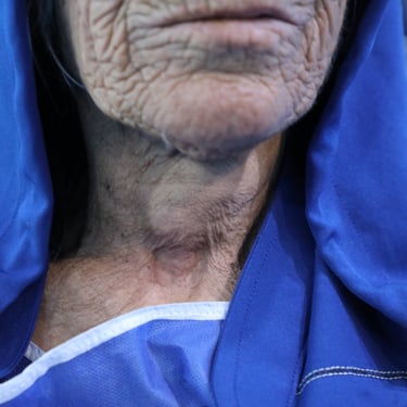
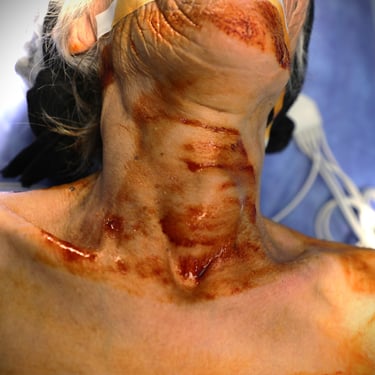
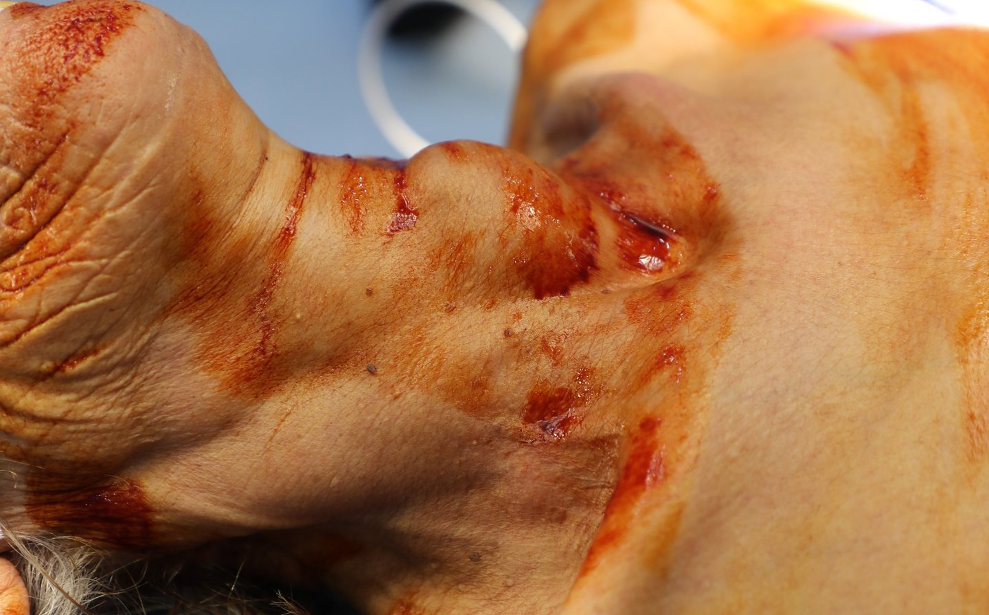
A 72-year-old female presented with an anterior neck lesion for 4 years. She has a long history of smoking and a past medical history of kidney stones and a heart condition. Her past surgical history is unremarkable, and she is not on any regular medication. There is no significant family history. On physical examination, the patient was found to have a Grade 3 goiter.
SURGERYHEAD AND NECK
4/12/20252 min read

Patient Information:
A 72-year-old female presented with an anterior neck lesion for 4 years. She has a long history of smoking and a past medical history of kidney stones and a heart condition. Her past surgical history is unremarkable, and she is not on any regular medication. There is no significant family history. On physical examination, the patient was found to have a Grade 3 goiter.
Laboratory Investigations:
Initial laboratory findings showed TSH levels of <0.005 uIU/mL, indicating thyrotoxicosis. Her Free Thyroxine (FT4) was 32.9 pmol/L, suggesting hyperthyroidism. Additionally, TSH Receptor Antibodies (TRAb) were elevated at 20.9 IU/L, and Anti-Thyroid Peroxidase Antibody (ATPO) was 14.8 IU/mL, further supporting thyroid dysfunction.
Ultrasound Findings:
Ultrasound of the neck revealed a normal right thyroid lobe (50 × 17 × 15 mm) with a small <10 mm TR3 nodule. The left thyroid lobe was enlarged (74 × 40 × 39 mm) due to a large, irregular, hypoechoic mass with heterogeneous echogenicity, extending into the left isthmus and showing increased vascularity. This mass was highly suspicious for malignancy.
Additionally, multiple bilateral cervical lymph nodes were identified, with the largest being 26 × 16 mm in the right group I and 25 × 10 mm in the left group III. These nodes had inhomogeneous texture and were mildly vascular, raising concern for metastasis. An irregularity was also noted in the inner surface of the thyroid cartilage, which was suspicious for thyroid invasion.
CT Findings:
A contrast-enhanced CT scan of the neck revealed a large thyroid mass with associated bilateral cervical lymphadenopathy. There was severe tracheal narrowing due to the thyroid mass, and the scan also showed multiple pulmonary nodules, liver lesions, and renal abnormalities, raising concern for metastatic spread.
Fine Needle Aspiration (FNA) Results:
Thyroid Mass: Atypia of undetermined significance (AUS), Bethesda III, which indicates a need for further diagnostic evaluation.
Cervical Lymph Node: FNA showed metastatic thyroid carcinoma.
Oncology Consultation:
The patient was referred to an oncologist, and the findings were suggestive of Hurthle cell carcinoma (a variant of follicular carcinoma), with pulmonary, liver, and renal lesions likely indicating metastatic disease. The oncologist recommended a liver biopsy and a total thyroidectomy with left cervical lymph node dissection as part of palliative treatment.
Surgical Intervention:
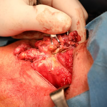
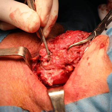
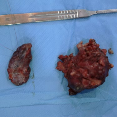
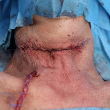
The patient underwent a total thyroidectomy, including a left cervical lymph node dissection. The surgery revealed that the left thyroid mass involved the recurrent laryngeal nerve (RLN) and adjacent muscles, and the mass had invaded the left trachea. The largest right lateral lymph node and the left lateral lymph nodes were removed.
Histopathological Examination (HPE):
Thyroid Histopathology: The tumor was diagnosed as anaplastic thyroid carcinoma, arising from a high-grade papillary thyroid carcinoma. The tumor was unifocal, with extensive necrosis, a high mitotic rate (>25 mitoses/2mm), and extensive lymphatic and vascular invasion. Perineural invasion was identified, and the tumor had minimal extra-thyroid extension into surrounding fibroadipose tissue and muscle. The tumor was at least 6 cm in size, and no tumor capsule was present. Diffuse thyroid hyperplasia was also noted.
Lymph Node Involvement: Four lymph nodes were involved with metastasis: one from the right side showing high-grade papillary carcinoma, and three from the left, showing both anaplastic and high-grade papillary carcinomas, with extranodal extension.
Additional Findings: The tumor showed papillary thyroid carcinoma with increased mitotic activities and necrosis, and underwent anaplastic transformation.
Postoperative Management:
The patient’s postoperative management focused on addressing the metastatic spread, with the involvement of pulmonary, liver, and renal lesions. Given the aggressive nature of the anaplastic carcinoma, a multidisciplinary approach was employed, including radiation therapy and chemotherapy. The prognosis remains poor, and the patient’s treatment is primarily palliative.
Gallery
