Cystic Nodule with Multinodular Goiter and Branchial Cleft Cyst
A 46-year-old female presented with painless right-sided neck swelling, which had been present for about one month. The patient had no significant past medical history, and there was no family history of thyroid cancer.
SURGERY
7/9/20242 min read

Case Presentation:
A 46-year-old female presented with painless right-sided neck swelling, which had been present for about one month. The patient had no significant past medical history, and there was no family history of thyroid cancer.
General Examination:
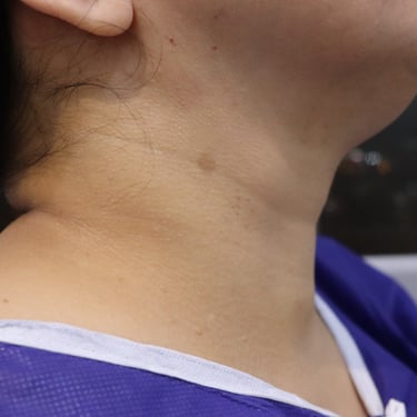
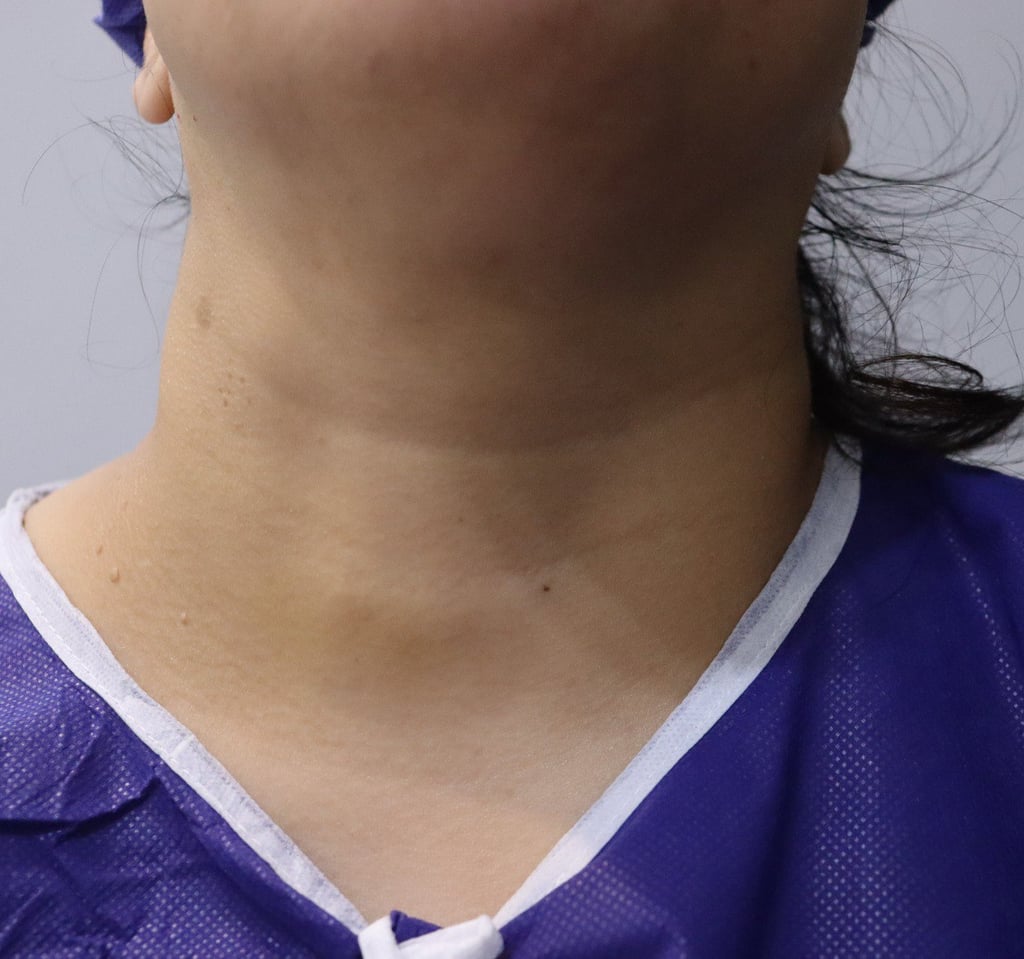
Inspection: A spherical mass on the right side of the neck, approximately 3 x 2 cm in size, with normal skin color.
Palpation: A firm, non-tender, non-pulsatile, and non-hot mass was palpable. The patient also had multinodular goiter (MNG).
Investigations: None
Laboratory Tests:
TSH: 0.02 µIU/mL
Free T4: 21.3 pmol/L
Serum Calcium: 9.6 mg/dL
Thyroglobulin: >4.8 ng/mL (normal range: 3.5–77 ng/mL)
Neck Ultrasound Findings:
Multinodular Goiter (MNG):
Right side TR3 nodule: 14 x 9 x 7 mm
Left side TR3 nodule: 18 x 16 x 19 mm
Right Neck:
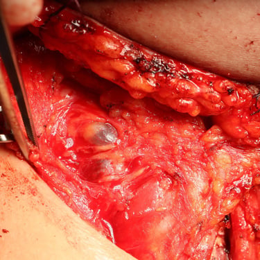
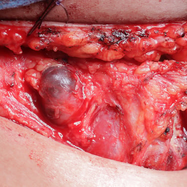
A highly suspicious complex cystic nodule measuring 29 x 18 x 15 mm located anterolaterally to the distal third of the right common carotid artery.
Cytology:
Thyroid Nodule: Negative for malignancy, benign follicular nodule.
Cervical Nodule: Cystic content, non-diagnostic.
Thyroglobulin from cystic content: >500 ng/mL
Surgery:
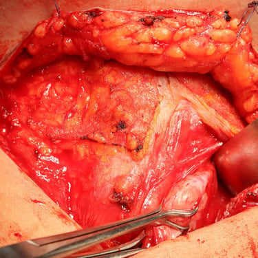
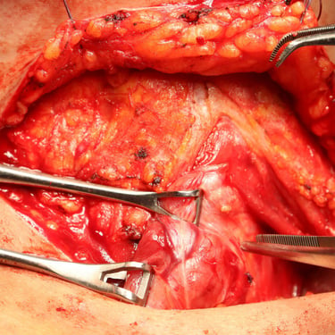
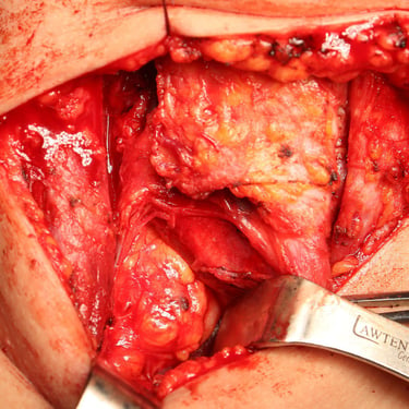
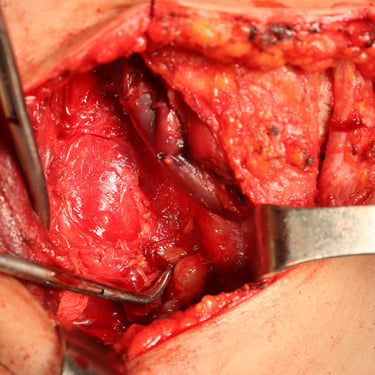
The patient underwent a total thyroidectomy along with right central and right lateral cervical lymph node dissection.
Gross Findings (Surgical Specimen):
Thyroidectomy:
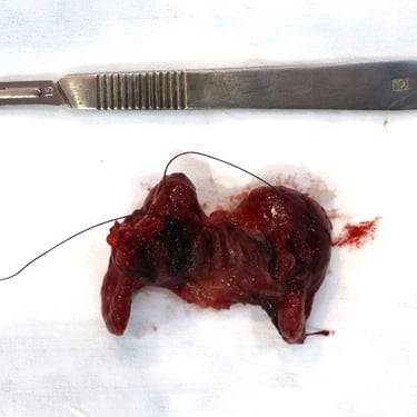
Right lobe: 5 x 4 x 2 cm
Left lobe: 4.5 x 3 x 1 cm
Isthmus: 2.5 x 1.5 x 1 cm
Multiple nodules of varying sizes, with hemorrhage and fibrosis present in the thyroid gland.
Right Central Lymph Nodes: 4 lymph nodes, the largest measuring 0.5 cm.
Right Lateral Lymph Nodes: 29 lymph nodes, with one uniloculated cyst (2.5 cm) filled with hemorrhagic gelatinous material and a 0.7 cm solid white component.
Histopathological Examination (HPE):
Thyroid Gland:
Follicular nodular disease with focal lymphocytic thyroiditis.
The thyroid tissue consisted of various-sized nodules composed of benign follicles lined by follicular epithelial cells and filled with colloid material, predominantly arranged as macrofollicles. Associated features included cystic hemorrhage, fibrosis, and focal lymphocytic infiltration forming lymphoid follicles with active germinal centers.
Right Lateral Cervical Cyst:
A thin cyst wall lined by a single layer of benign epithelial cells. The cyst contained abundant benign lymphoid cells forming lymphoid follicles with active germinal centers. The lumen was filled with eosinophilic secretion and a few inflammatory cells.
No evidence of thyroid tissue or metastatic thyroid carcinoma in the cyst.
Lymph Nodes:
Benign with reactive follicular hyperplasia. The architecture was preserved.
Diagnosis:
Thyroid: Follicular nodular disease with focal lymphocytic thyroiditis.
Right Lateral Cervical Cyst: Branchial cleft cyst.
Cervical Lymph Nodes: Benign lymph nodes with reactive follicular hyperplasia.
No malignancy: No primary thyroid carcinoma or metastatic thyroid carcinoma was detected in the thyroid gland, cervical lymph nodes, or cyst.
Comment:
There was no evidence of thyroid carcinoma either grossly or microscopically in the thyroid gland.
No benign thyroid tissue or metastatic thyroid carcinoma was found in the cervical lymph nodes or lateral cervical cyst.
Conclusion:
The case represents a benign thyroid condition with a complex cystic mass in the cervical region, later identified as a branchial cleft cyst. The histopathological examination confirmed the diagnosis of follicular nodular disease with focal lymphocytic thyroiditis and reactive lymph nodes. No evidence of malignancy was present in any of the tissue samples, and the patient is expected to recover without further complications.
Gallery

