Exophytic Thyroid Mass Mimicking Paraganglioma in a 48-Year-Old Female
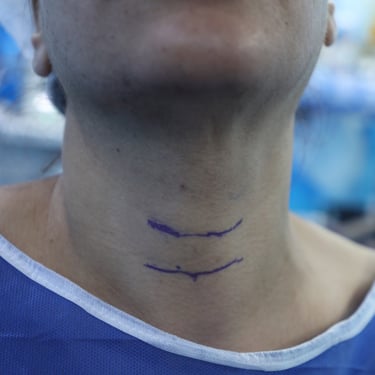
A 48-year-old female presented with a left-sided neck swelling noted incidentally, without associated symptoms such as pain, dysphagia, voice changes, or systemic complaints. On examination, the swelling was firm, round, and located on the left side of the neck. There were no signs of inflammation or tenderness.
SURGERYHEAD AND NECKVIDEO
5/17/20252 min read


Patient Presentation:
A 48-year-old female presented with a left-sided neck swelling noted incidentally, without associated symptoms such as pain, dysphagia, voice changes, or systemic complaints. On examination, the swelling was firm, round, and located on the left side of the neck. There were no signs of inflammation or tenderness.
Medical and Surgical History:
Her past medical and surgical history was unremarkable, with no history of thyroid disease, radiation exposure, or previous surgeries. She denied any current medication use, and her family history was negative for thyroid or endocrine conditions.
Laboratory Investigations:
Thyroid function tests were within normal limits: TSH was 1.77 uIU/mL and Free T4 was 16.0 pmol/L. Autoimmune markers were also normal with Anti-TPO at 17.2 IU/mL and TRAb <0.800 IU/L. Inflammatory markers and calcium metabolism parameters were normal, including CRP (0.191 mg/L), PTH (41.7 pg/mL), and serum calcium (9.27 mg/dL).
Neck Ultrasound:
Ultrasound revealed both thyroid lobes had homogeneous echotexture. A tiny 3 mm micronodule was noted in the right lobe. On the left side, there was a well-defined, regular, predominantly solid exophytic nodule arising from the lower third of the lobe, measuring 45 × 36 × 29 mm, with retrosternal extension and increased perinodular and intranodular vascularity. No calcifications were seen. The lesion was categorized as TIRADS 3.
CT Neck and Upper Chest with Contrast:
CT imaging showed a well-defined, smooth, hypervascular solid mass with a non-enhancing cystic/necrotic center located posteroinferior to the left thyroid gland, measuring approximately 4.7 × 4.2 × 3.0 cm. It displaced the thyroid anteriorly and caused mild deviation of the trachea and esophagus to the right. Although the lesion had no definitive connection to the thyroid, its density was similar to thyroid parenchyma on both arterial and venous phases, raising the possibility of an exophytic thyroid nodule. Differential diagnosis included a thyroid-associated paraganglioma, or less likely, a parathyroid mass. No pathologic cervical lymphadenopathy was identified.
Fine-Needle Aspiration (FNA):
An FNA of the lesion was performed and interpreted as Bethesda II, consistent with a benign follicular nodule, and no evidence of malignancy or paraganglioma.
Surgical Management:
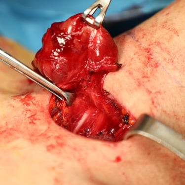
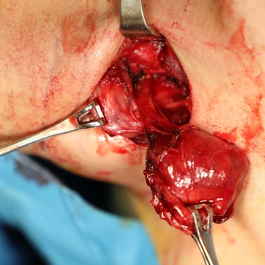
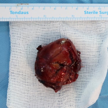
Despite the benign FNA, due to the lesion's size, retrosternal extension, hypervascularity, and uncertain origin, the patient was scheduled for surgical removal. She underwent excision of the left thyroid lobe mass with a working diagnosis of either an exophytic thyroid nodule or thyroid-associated paraganglioma.
Histopathology Report:
Histopathological analysis confirmed the diagnosis of an adenomatoid thyroid follicular nodule. There was no evidence of malignancy, paraganglioma, or parathyroid pathology. The findings supported a benign exophytic thyroid nodule as the final diagnosis.
Head And Neck Surgeon: Dr.Abdulwahid M. Salih
Co-Surgeon : Dr Yadgar Abdulhameed Saeed
Assistant : Dr. Abdullah Osman Hassan

