Hemangioma in a 16-Year-Old Female
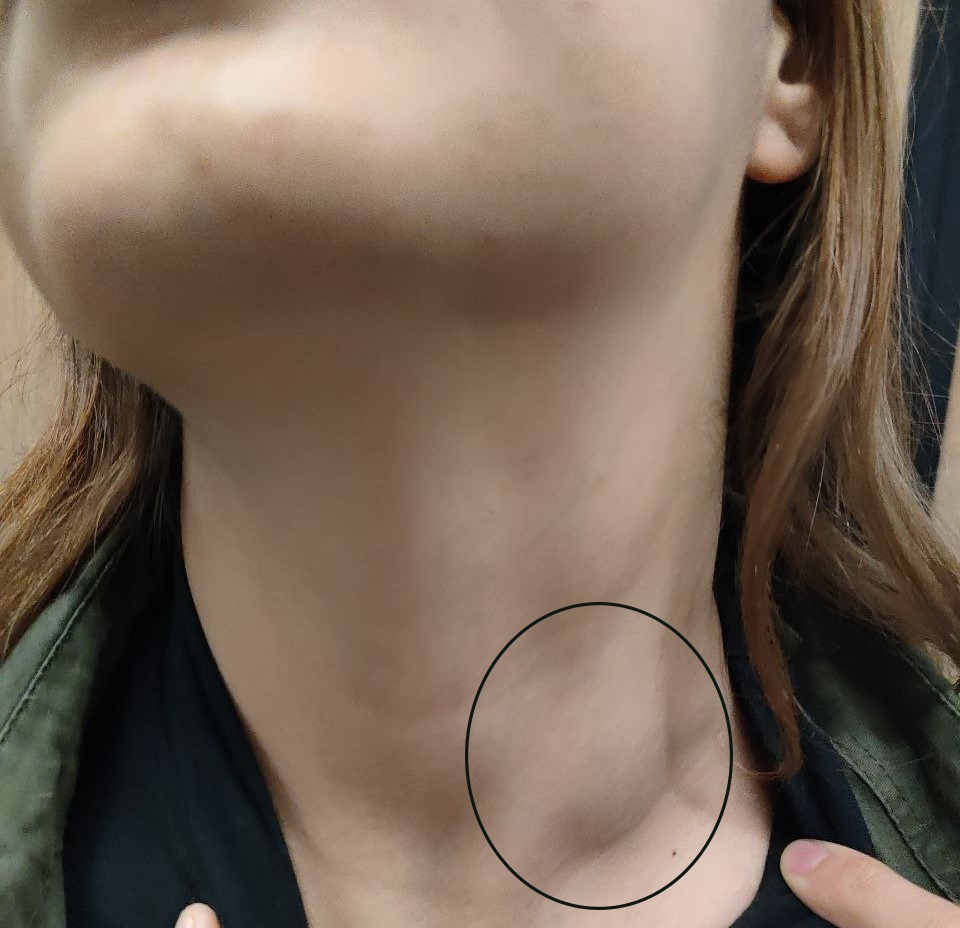
A 16-year-old female presented with a left-sided neck swelling that had been progressively increasing in size over two years. She reported no associated symptoms, and her past medical and surgical history was unremarkable. The swelling was noted to enlarge during straining.
SURGERY
2/10/20241 min read


Case Presentation:
A 16-year-old female presented with a left-sided neck swelling that had been progressively increasing in size over two years. She reported no associated symptoms, and her past medical and surgical history was unremarkable. The swelling was noted to enlarge during straining.
Clinical Examination:
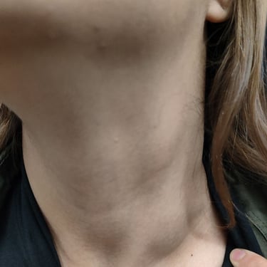
On examination, a soft, compressible lump was observed on the left side of the neck. The mass was non-tender and demonstrated dynamic enlargement with increased intrathoracic pressure. No signs of inflammation or skin discoloration were present.
Imaging Findings:
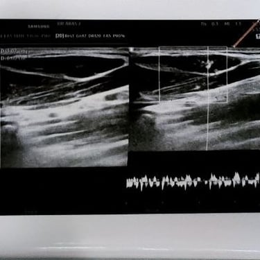
Neck ultrasound revealed a well-defined, heterogeneously hypoechoic vascular lesion measuring 30x20x6 mm, located in the lower third of the left sternocleidomastoid muscle and subcutaneous tissue. The lesion was mildly vascular on color Doppler, with characteristics suggestive of a hemangioma. Additionally, a few small lymph nodes were noted in the upper left neck, with the largest (18x6 mm) in the submandibular region, appearing inflammatory in nature.
Laboratory Findings:
Blood tests, including thyroid function tests (TSH: 2.66 uIU/ml, FT4: 14.4 pmol/L, T3: 1.60 ng/ml), inflammatory markers (CRP: 2.66 mg/L, ESR: 10 mm/hr), and complete blood count (CBC), were within normal limits, except for a mildly decreased hemoglobin (HGB: 9.9 g/dl) and hematocrit (HCT: 31.2%).
Management Plan:
Following a multidisciplinary team (MDT) discussion, reassurance and regular follow-up were recommended for the patient. Given the benign nature of the lesion and the absence of significant symptoms or complications, conservative management was deemed appropriate.
Image Gallery:


