High-Grade Adenocarcinoma of the Parotid Gland with Cervical Lymph Node Metastasis
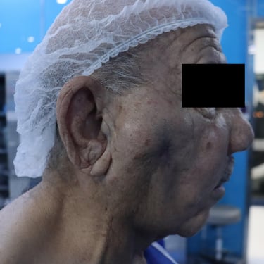
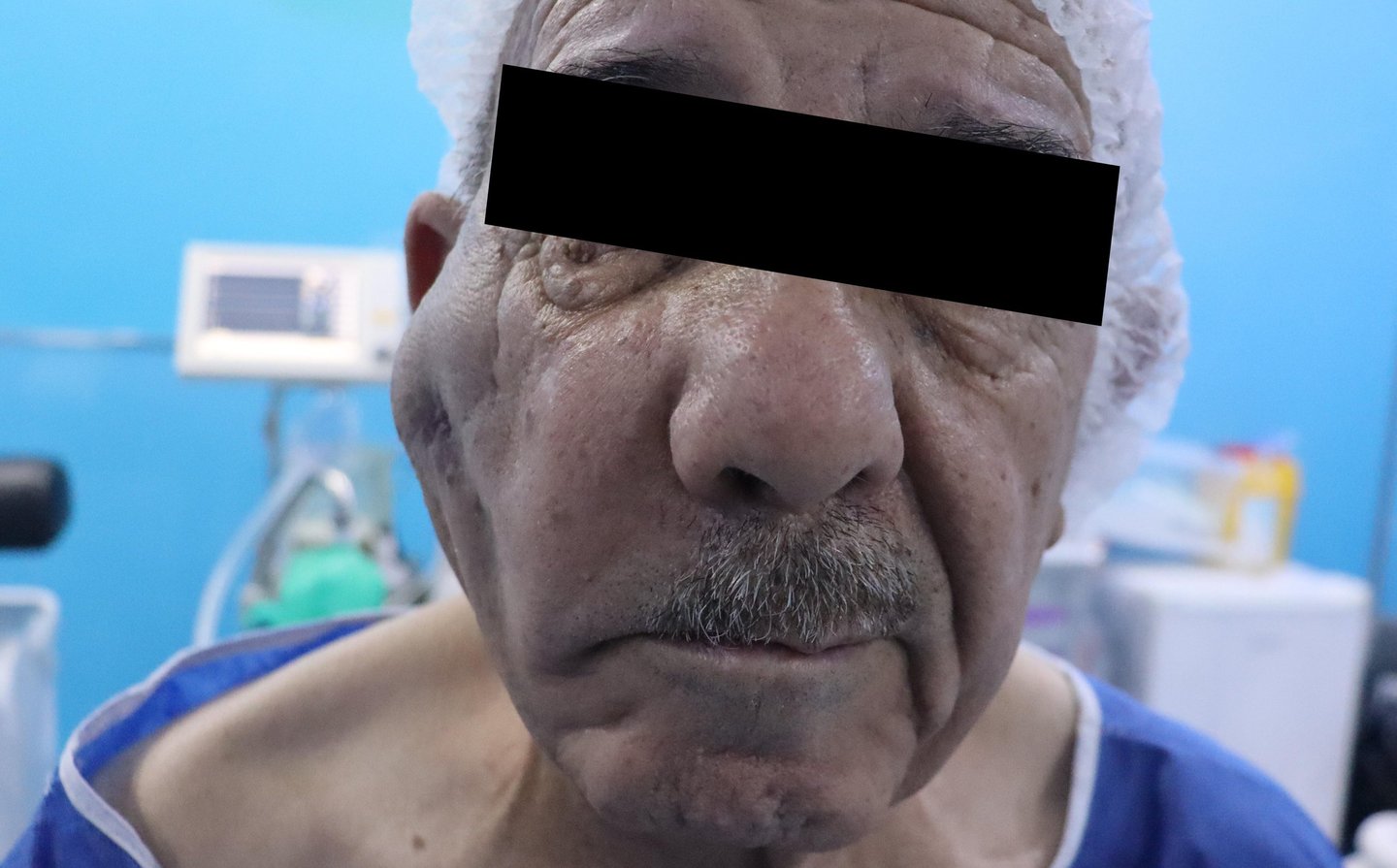
A 79-year-old male presented with a right preauricular swelling persisting for 40 years. Over the last few years, the swelling had progressively enlarged, leading to facial asymmetry. The patient had no history of smoking and no significant past medical or surgical history. On examination, the mass was hard and movable.
SURGERYHEAD AND NECK
2/20/20252 min read

Chief Complaint:
A 79-year-old male presented with a right preauricular swelling persisting for 40 years. Over the last few years, the swelling had progressively enlarged, leading to facial asymmetry. The patient had no history of smoking and no significant past medical or surgical history. On examination, the mass was hard and movable.
Multidisciplinary Team (MDT) Discussion and Oncologist Referral:
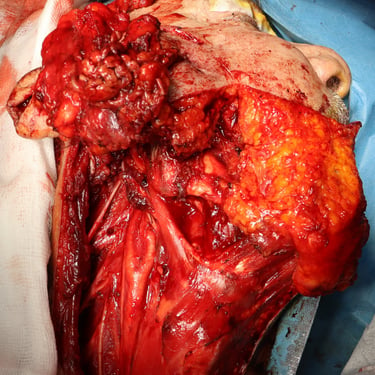
Given the clinical suspicion of carcinoma ex-pleomorphic adenoma, the case was discussed in an MDT meeting. A referral was made to an oncologist to determine whether surgical intervention was feasible or if the patient required chemotherapy and radiation. Based on the oncologist’s recommendation, the patient was scheduled for total parotidectomy with preservation of the upper branch of the facial nerve, along with a radical right neck dissection.
Investigations:
Routine laboratory investigations revealed a serum blood urea level of 32.5 mg/dL and a creatinine level of 1.14 mg/dL. Viral screening was negative. Preoperative fine-needle aspiration (FNA) suggested a poorly differentiated carcinoma, with suspicion of carcinoma ex-pleomorphic adenoma and metastatic right cervical lymph node involvement.
Surgical Procedure and Pathological Findings:
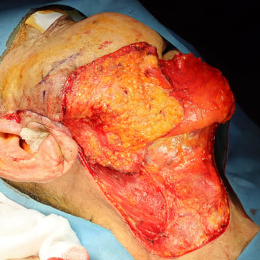
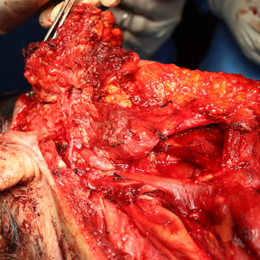
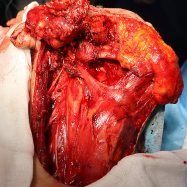
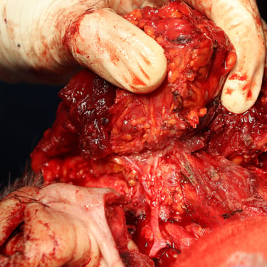
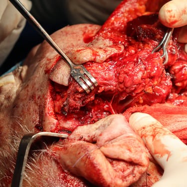
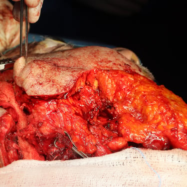
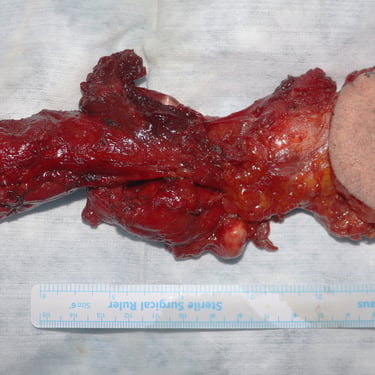
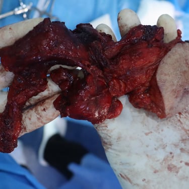
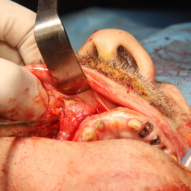
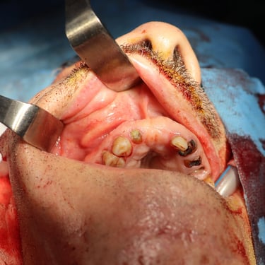
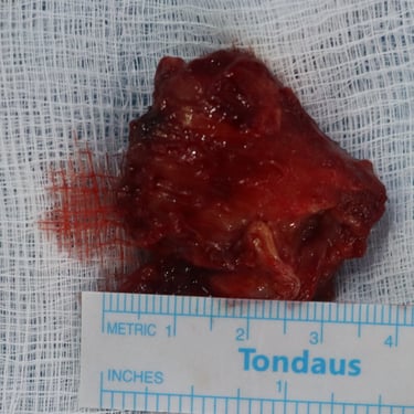
The patient underwent total parotidectomy with radical right neck dissection and excision of a mass from the canine space. Histopathological examination confirmed a high-grade adenocarcinoma of no specific type with extensive perineural and lymphovascular invasion. The tumor measured 6 cm with an infiltrative border and involved the deep resection margin. Skin involvement was noted, though without ulceration.
Seventeen out of 42 dissected lymph nodes showed macrometastases with extranodal extension, the largest measuring 2 cm. The pathological staging was determined as pT4a N3b R1 (AJCC 8th edition). Additional findings included chronic fibrosing sialadenitis and extensive fibrosis.
Immunohistochemistry and Further Recommendations:
Immunohistochemical staining of the canine space mass indicated an atypical B-cell proliferation. The staining pattern did not confirm follicular lymphoma, prompting recommendations for further lymph node assessment and possible biopsy of any suspicious nodes elsewhere in the body.
Postoperative Course and Follow-up:
The patient had a good post-operative recovery with only transient facial asymmetry. He remained stable during the two-month follow-up period and was referred to an oncologist for further evaluation regarding the need for chemotherapy and radiotherapy.
Gallery

