Hodgkin’s Lymphoma in a 27-Year-Old Female
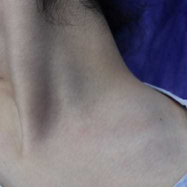
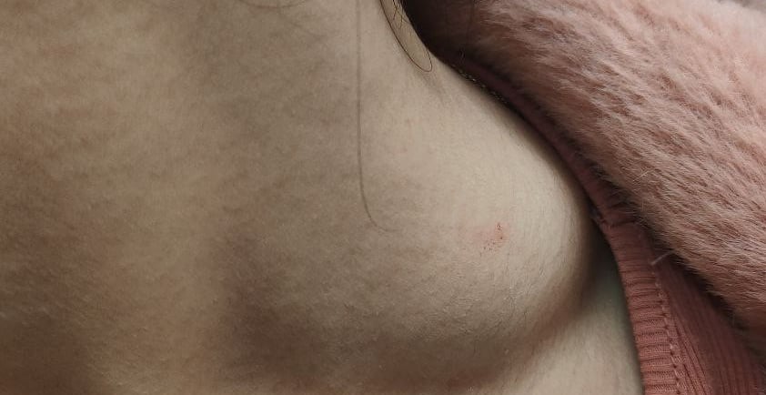
A 27-year-old female presented with a left-sided neck swelling that had been progressively enlarging over one month. She had no associated symptoms, and her past medical, surgical, and drug histories were unremarkable.
HEAD AND NECK
2/21/20241 min read

Case Presentation:
A 27-year-old female presented with a left-sided neck swelling that had been progressively enlarging over one month. She had no associated symptoms, and her past medical, surgical, and drug histories were unremarkable.
Laboratory Findings:
Blood tests showed a slightly elevated erythrocyte sedimentation rate (ESR) of 32 mm/hr (normal: 0–30 mm/hr) and an elevated C-reactive protein (CRP) level of 8.29 mg/L (normal: <5 mg/dL). Complete blood count (CBC), lactate dehydrogenase (LDH), and thyroid-stimulating hormone (TSH) levels were within normal ranges.
Imaging Findings:
Neck ultrasound revealed a highly suspicious, large, solid, hypoechoic, and mildly vascular lymph node measuring 42x25x18 mm in the left supraclavicular region with loss of the hilum. Smaller lymph nodes were noted in the right supraclavicular area (largest 17x8 mm) and in bilateral cervical levels III and IV, with loss of normal echo texture and cortical thickening. Abdominal imaging showed no significant lymphadenopathy, and the liver and spleen appeared normal. The thyroid, submandibular, and parotid glands were unremarkable.
Diagnostic and Treatment Approach:
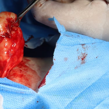
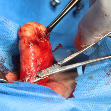
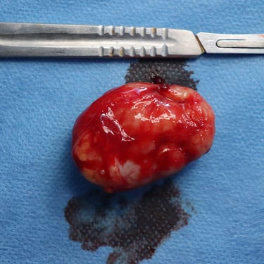
A multidisciplinary team (MDT) discussion led to the decision to perform an excisional biopsy of the largest lymph node for histopathological examination to establish a definitive diagnosis.
Histopathological and Immunohistochemical Findings:
The excised lymph node exhibited near-total effacement of its architecture with fibrous bands and sclerosis, consistent with nodular sclerosing classical Hodgkin’s lymphoma. Large, atypical mono- and multinucleated cells were present in a polymorphous background with numerous histiocytes, small lymphocytes, plasma cells, and eosinophils. Immunohistochemistry confirmed the diagnosis with CD15 and CD30 positivity in the large atypical cells, characteristic of classical Hodgkin’s lymphoma.
Conclusion:
The patient was diagnosed with nodular sclerosing classical Hodgkin’s lymphoma, confirmed by histopathology and immunohistochemistry. Further management was planned in consultation with oncology specialists for staging and initiation of appropriate therapy.
Image Gallery:
