Hypothyroidism and Ectopic Thyroid Tissue in a 22-Year-Old Female
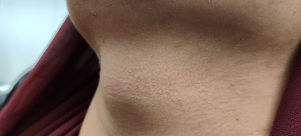
A 22-year-old female presented with an anterior neck mass of unknown duration. She had no known history of thyroid dysfunction before presentation.
SURGERY
12/4/20231 min read


Case Presentation:
A 22-year-old female presented with an anterior neck mass of unknown duration. She had no known history of thyroid dysfunction before presentation.
Laboratory Investigations:
Laboratory tests revealed elevated thyroid-stimulating hormone (TSH) levels with low free thyroxine (FT4) and triiodothyronine (T3) levels, indicating hypothyroidism. Anti-thyroid peroxidase antibody (ATPO) levels were within the normal range.
TSH: 19 uIU/ml (elevated)
FT4: 6.7 pmol/L (low)
T3: 1.04 ng/ml (low)
ATPO: 12 IU/ml (normal)
Imaging Findings:
Neck ultrasound showed the absence of a thyroid gland in its normal anatomical position. Instead, a well-defined complex ectopic thyroid tissue was identified in the midline of the neck, measuring 42 × 32 × 19 mm.
Diagnosis:
The findings confirmed hypothyroidism due to ectopic thyroid tissue in the midline of the neck.
Management Plan:
The patient was started on thyroid hormone replacement therapy with thyroxine 100 mcg once daily to correct hypothyroidism and prevent further glandular hypertrophy. She was scheduled for regular follow-up to monitor thyroid function, hormone levels, and any changes in the size or function of the ectopic thyroid tissue.
Image Gallery


