Incidental Finding of Anaplastic Thyroid Carcinoma in a 67-Year-Old Female
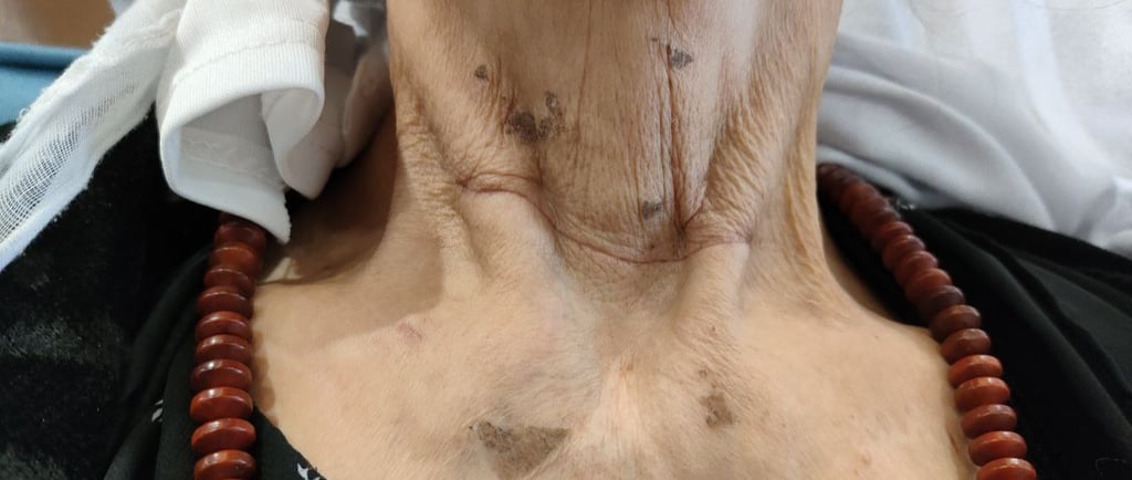
A 67-year-old female presented with persistent anterior neck swelling for the past 10 years. Her medical history was notable for diabetes, hypertension, and hyperthyroidism. Preoperative investigation revealed low thyroid-stimulating hormone (TSH) levels and elevated free thyroxine (FT4) levels, indicative of hyperthyroidism. The calcium level was within normal limits. Upon physical examination, a large goiter was visibly prominent.
SURGERY
12/1/20231 min read


Case Presentation:
A 67-year-old female presented with persistent anterior neck swelling for the past 10 years. Her medical history was notable for diabetes, hypertension, and hyperthyroidism. Preoperative investigation revealed low thyroid-stimulating hormone (TSH) levels and elevated free thyroxine (FT4) levels, indicative of hyperthyroidism. The calcium level was within normal limits. Upon physical examination, a large goiter was visibly prominent.
TSH Levels: 0.005 (low)
FT4 Levels: 27.04 (elevated)
Calcium Levels: 9.04 (normal)
Imaging Findings:
Neck ultrasound revealed a multinodular goiter with the following measurements:
Right lobe: 77 × 47 × 39 mm
Left lobe: 100 × 66 × 49 mm
Isthmus: 15 mm thick
The largest nodules were in the right lobe (37 × 31 × 30 mm, TR3) and left lobe (59 × 40 × 38 mm, TR3), with macrocalcification present. A mild left-sided retrosternal extension was also noted.
Surgical Management:
The patient underwent total thyroidectomy to treat the toxic multinodular goiter, as well as to address any potential malignancy.
Histopathological Examination:
Post-surgical examination revealed anaplastic thyroid carcinoma within a hyperplastic nodule. The tumor was characterized by high-grade, poorly differentiated squamous cell carcinoma, measuring 2.7 cm in size. It was localized to the right lobe without capsule infiltration. A single parathyroid gland attached to the left lobe was free from the tumor. No lymph node sampling was performed. According to the AJCC 8th edition, the tumor was staged as pT2 Nx.
Post-Operative and Follow-Up:
Following surgery, the patient’s serum calcium levels were recorded at 8.4. She has been referred to an oncologist for further management and surveillance, and she is currently doing well post-operatively.


