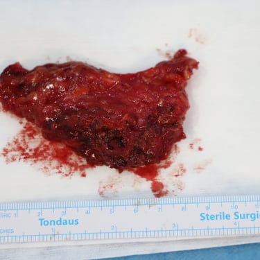
Left Infra-Auricular Mass Diagnosed as Pleomorphic Adenoma
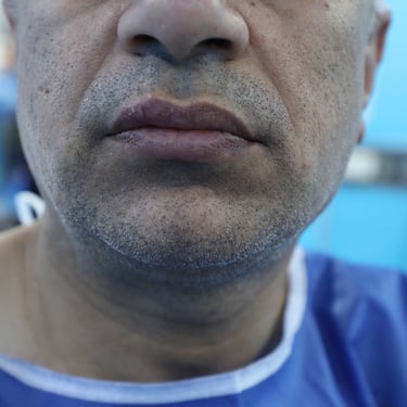
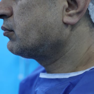
A 43-year-old male smoker presented with a left infra-auricular mass persisting for three years. His past medical and surgical history was unremarkable.
SURGERYHEAD AND NECKVIDEO
5/27/20251 min read


Patient Presentation:
A 43-year-old male smoker presented with a left infra-auricular mass persisting for three years. His past medical and surgical history was unremarkable.
Laboratory Investigations:
Blood investigations revealed a blood urea level of 22.0 mg/dL, serum creatinine of 1.01 mg/dL, and serum glucose of 132 mg/dL.
Ultrasound Findings:
Ultrasound of the left parotid gland showed a well-defined, oval, hypoechoic, and hypovascular lesion with posterior enhancement, located in the superficial lobe of the left parotid, measuring 20 × 11 mm. The findings were suggestive of pleomorphic adenoma. Multiple benign-appearing lymph nodes were noted on both sides, predominantly on the left, with the largest measuring 20 × 10 mm. The right parotid and bilateral submandibular salivary glands were normal. The thyroid gland was also normal, with no detected lesions.
Biopsy and Diagnosis:
A tru-cut biopsy of the left parotid lesion confirmed the diagnosis of pleomorphic adenoma.
Surgical Intervention:
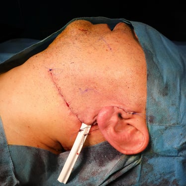
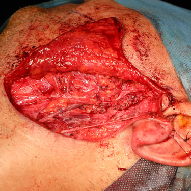
The patient underwent a left superficial parotidectomy with the excision of two large cervical lymph nodes.
Histopathological Examination (HPE):
Histopathology confirmed a diagnosis of pleomorphic adenoma (benign mixed tumor of the salivary gland). The excised lymph nodes showed reactive follicular hyperplasia with no evidence of malignancy.
Postoperative Outcome:
The patient remained well during follow-up, with no evidence of facial palsy or other complications.

