Left Preauricular Basal Cell Carcinoma
A 69-year-old male presented with a left preauricular lesion that had been slowly increasing in size over the past year. The patient reported no associated symptoms such as bleeding, itching, or discharge. He had no significant past medical or surgical history.
SURGERY
6/4/20242 min read

Case Presentation:
A 69-year-old male presented with a left preauricular lesion that had been slowly increasing in size over the past year. The patient reported no associated symptoms such as bleeding, itching, or discharge. He had no significant past medical or surgical history.
Clinical Examination:
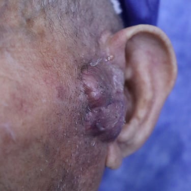
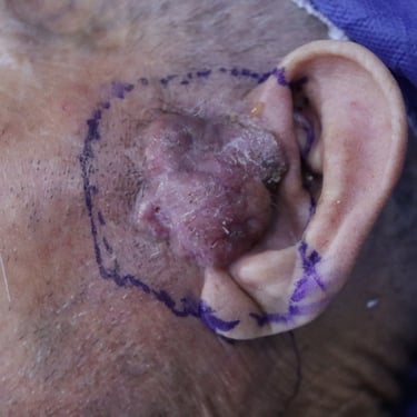
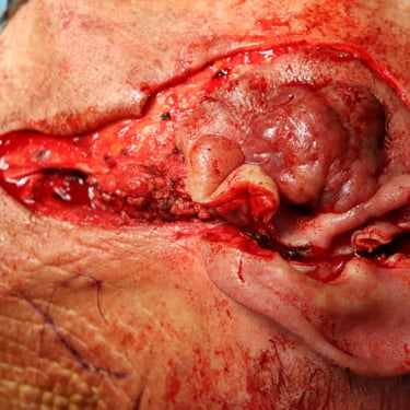
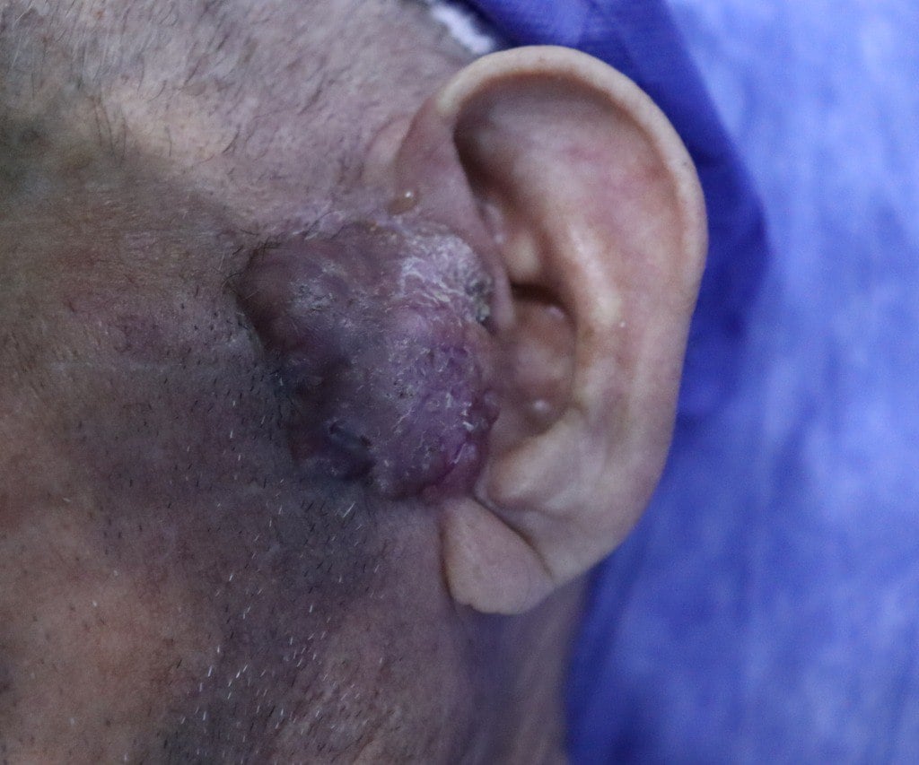
On inspection, the lesion appeared as a solitary, well-lobulated, red-colored mass measuring approximately 4 cm in length and 1 cm in width. There was no surrounding skin ulceration or significant redness. On palpation, the mass was firm, irregular, mobile, non-tender, and not warm to the touch.
Imaging Findings:
Ultrasound of the left preauricular region showed a solid, hypoechoic, hypervascular mass measuring 4x3x1.8 cm involving the skin and subcutaneous tissue, with partial involvement of the external ear. A few small, non-suspicious lymph nodes were noted under the mass and in the parotid group. Additionally, an incidental TR4 thyroid nodule was detected.
A head and neck MRI revealed a 3x1.4 cm skin mass anterior to the left auricle and posterior-lateral to the left cheek, without evidence of deep invasion.
Biopsy and Cytology:
An incisional biopsy of the preauricular lesion confirmed basal cell carcinoma (BCC), nodular variant. Fine needle aspiration cytology (FNAC) of the thyroid nodule was negative for malignancy (Bethesda II).
Surgical Management:
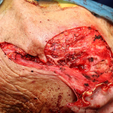
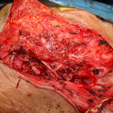
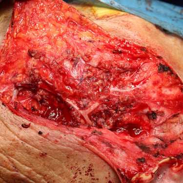
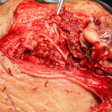
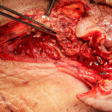
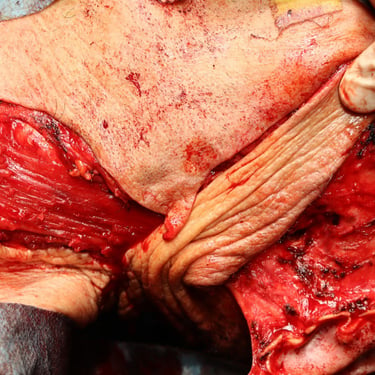
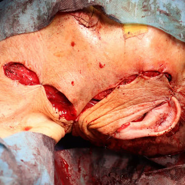
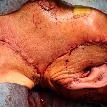
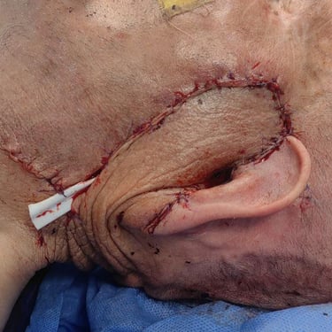
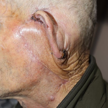
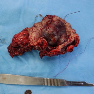
The patient underwent a wide local excision of the mass along with a superficial parotidectomy. During the procedure, all branches of the facial nerve were carefully explored and preserved. A rotational flap was used for reconstruction following tumor excision.
Histopathological Examination:
The histopathological analysis confirmed a nodular-type basal cell carcinoma measuring 3.5 cm in size. The tumor invaded beyond the cartilage and was adherent to the surface of the parotid gland. The surgical margins were clear, with a 3 mm and 8 mm clearance from the deep margin. No lymph node involvement was identified, and the parotid gland was unremarkable.
Postoperative Management:
Following surgery, the patient was scheduled for adjuvant radiotherapy to ensure local disease control and minimize the risk of recurrence.
Conclusion:
This case highlights an advanced basal cell carcinoma with extension beyond the skin, requiring wide excision and parotid gland preservation. Despite its locally aggressive nature, the tumor was successfully managed with surgery, and the patient was referred for further radiotherapy to reduce the chance of recurrence.
Image Gallery:

