Long-Standing Neck Swelling Due to Multinodular Goiter with Retrosternal Extension
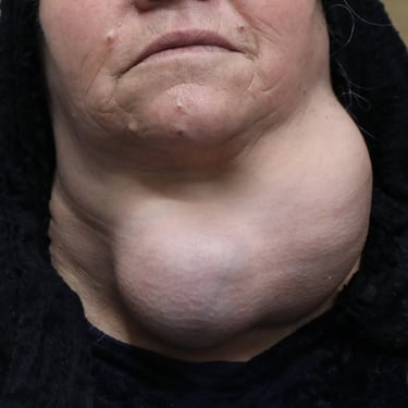
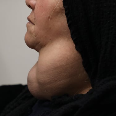
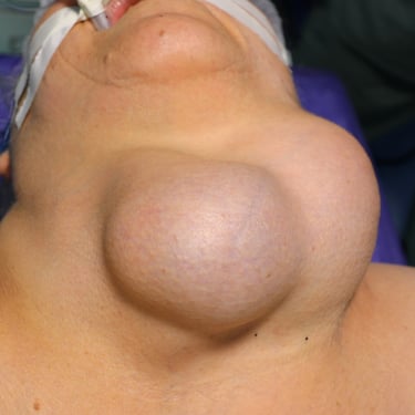
A 62-year-old female presented with a long-standing neck swelling that had been progressively increasing in size for more than 40 years. She is a non-smoker and has a history of hypertension.
SURGERYHEAD AND NECKVIDEO
4/30/20252 min read


Patient Presentation:
A 62-year-old female presented with a long-standing neck swelling that had been progressively increasing in size for more than 40 years. She is a non-smoker and has a history of hypertension.
Laboratory Investigations:
Laboratory tests revealed a TSH level of 0.407 uIU/mL and a Free Thyroxine (FT4) level of 9.70 pmol/L. Anti-Thyroid Peroxidase Antibody (Anti-TPO) was 12.9 IU/mL. Thyroglobulin (Tg) was significantly elevated at 205 ng/mL. Serum calcium was measured at 9.14 mg/dL, and calcitonin was 29.94 pg/mL.
Thyroid Ultrasound Findings:
Ultrasound examination showed a markedly enlarged thyroid gland with a heterogeneous echotexture and multiple well-defined nodules of varying sizes. The right lobe measured 120 × 67 × 59 mm, while the left lobe was significantly enlarged and could not be fully measured. The isthmus thickness was 34 mm. The largest nodule in the right lobe measured 55 × 45 × 37 mm (TR3), while the largest nodule in the left lobe measured 115 × 100 × 87 mm (TR3). Mildly increased vascularity was observed, along with macrocalcifications within some nodules. There was evidence of retrosternal extension, with the thyroid extending throughout the neck and significantly compressing the surrounding tissues. The findings were suggestive of multinodular goiter (MNG).
Neck and Upper Chest CT Findings:
Contrast-enhanced CT of the neck and upper chest confirmed heterogeneous nodular goiter with bilateral thyroid lobe enlargement. The right lobe measured 11 × 6 cm, extending posterior to the hypopharynx into the prevertebral space at C2–C4 levels, crossing midline, with mild retrosternal extension. The left lobe measured 14 × 10 cm with a significant retrosternal component. Two separate midline nodules were identified: one arising from the isthmus measuring 7 × 4 cm and another superficial subcutaneous nodule measuring 7 × 6 cm. Mass effect from the enlarged thyroid was causing mild upper tracheal luminal narrowing.
Surgical Intervention and Outcome:
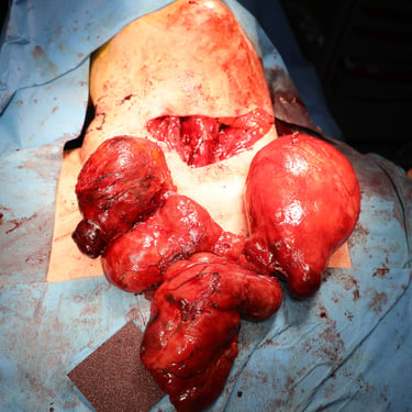
The patient underwent a total thyroidectomy. Histopathological examination (HPE) confirmed the presence of thyroid follicular nodular disease. On postoperative follow-up, the patient remained clinically stable, with no voice changes or significant complications.
Head and Neck Surgeon: Prof. Dr. Abdulwahid Mohammed Salih
Co-surgeon: Dr. Aso Saeed Muhialdeen & Dr. Muhamad Bag
Assistant: Dr. Harun Amanj Ahmed & Dr. Dindar Hussein Hama

