Management of a 22-Year-Old Male with a Swelling in the Right Post-Auricular Region
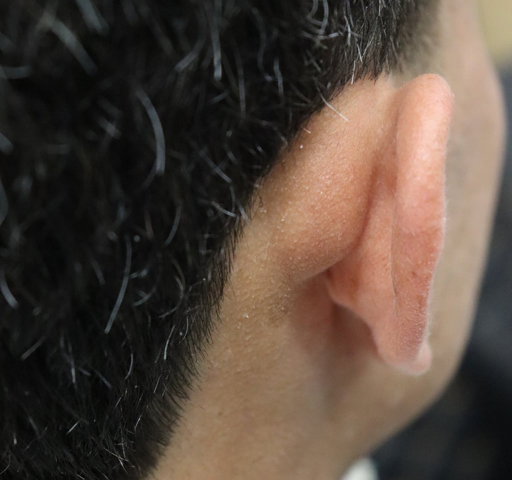
A 22-year-old male presented with a swelling in the right post-auricular region, which he had noticed several months prior. The mass was asymptomatic, with no associated pain, discharge, or noticeable changes in size. There were no identifiable factors that exacerbated or alleviated the condition. The patient had no significant past medical, surgical, or family history.
SURGERY
8/6/20242 min read


Case Presentation:
A 22-year-old male presented with a swelling in the right post-auricular region, which he had noticed several months prior. The mass was asymptomatic, with no associated pain, discharge, or noticeable changes in size. There were no identifiable factors that exacerbated or alleviated the condition. The patient had no significant past medical, surgical, or family history.
Physical Examination:
General Examination:
The patient appeared well-nourished and in no acute distress. Vital signs were stable and within normal limits.
Inspection:
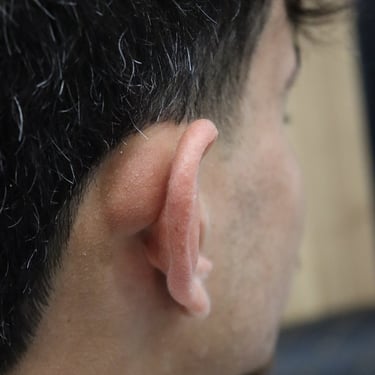
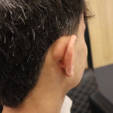
A lump measuring approximately 2 × 2 × 1 cm was identified in the right post-auricular region.
Palpation:
The lump was soft, non-tender, and fluctuating upon palpation. No palpable cervical lymph nodes were noted.
Imaging Studies:
Ultrasound of the Neck:
A well-defined, thin-walled (1 mm) cystic lesion was identified in the right post-auricular region, measuring 28 × 20 × 9 mm.
The lesion was hypovascular on color Doppler. The surrounding tissue appeared normal, suggesting a dermoid or epidermoid cyst.
Surgical Intervention:
The patient underwent surgical excision of the right post-auricular mass. The mass was excised without complication, and the tissue was sent for histopathological examination.
Post-Operative Status:
The patient was vitally stable after surgery and was discharged on the same day as the operation.
Histopathological Examination (HPE):
The histopathology report revealed a ruptured epidermoid inclusion cyst with a giant cell reaction and inflammation. No malignancy was detected.
Discussion:
This case represents a typical presentation of a dermoid or epidermoid cyst, which is a common benign lesion of the skin. These cysts are often slow-growing and asymptomatic, as seen in this patient. The swelling is usually soft, non-tender, and may fluctuate, which was consistent with the physical examination findings.
The diagnosis is often confirmed with imaging studies such as ultrasound, which can help delineate the cystic nature of the lesion and guide the surgical approach. In this case, the ultrasound suggested a dermoid or epidermoid cyst, both of which are typically benign lesions. FNAC or histopathology after surgical excision remains important for confirming the diagnosis and ruling out malignancy.
Surgical excision is the treatment of choice for symptomatic or bothersome cysts. This patient had an uncomplicated recovery, and histopathology confirmed the benign nature of the lesion with no evidence of malignancy.
Conclusion:
This case highlights the importance of clinical examination and imaging in diagnosing benign cystic lesions of the head and neck. Surgical excision is effective for managing these lesions, and post-operative follow-up is generally straightforward, with a good prognosis. Regular monitoring and awareness of potential complications such as infection or rupture are crucial.
Gallery


