Management of a 40-Year-Old Male with an Infected Second Branchial Cleft Cyst
A 40-year-old male patient presents with a right submandibular mass that has been present for the past two months.
SURGERYHEAD AND NECK
8/24/20242 min read

Chief Complaint:
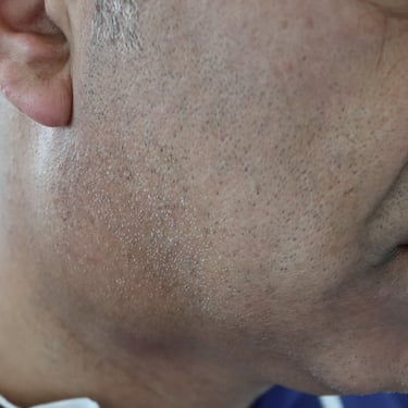
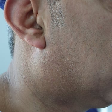
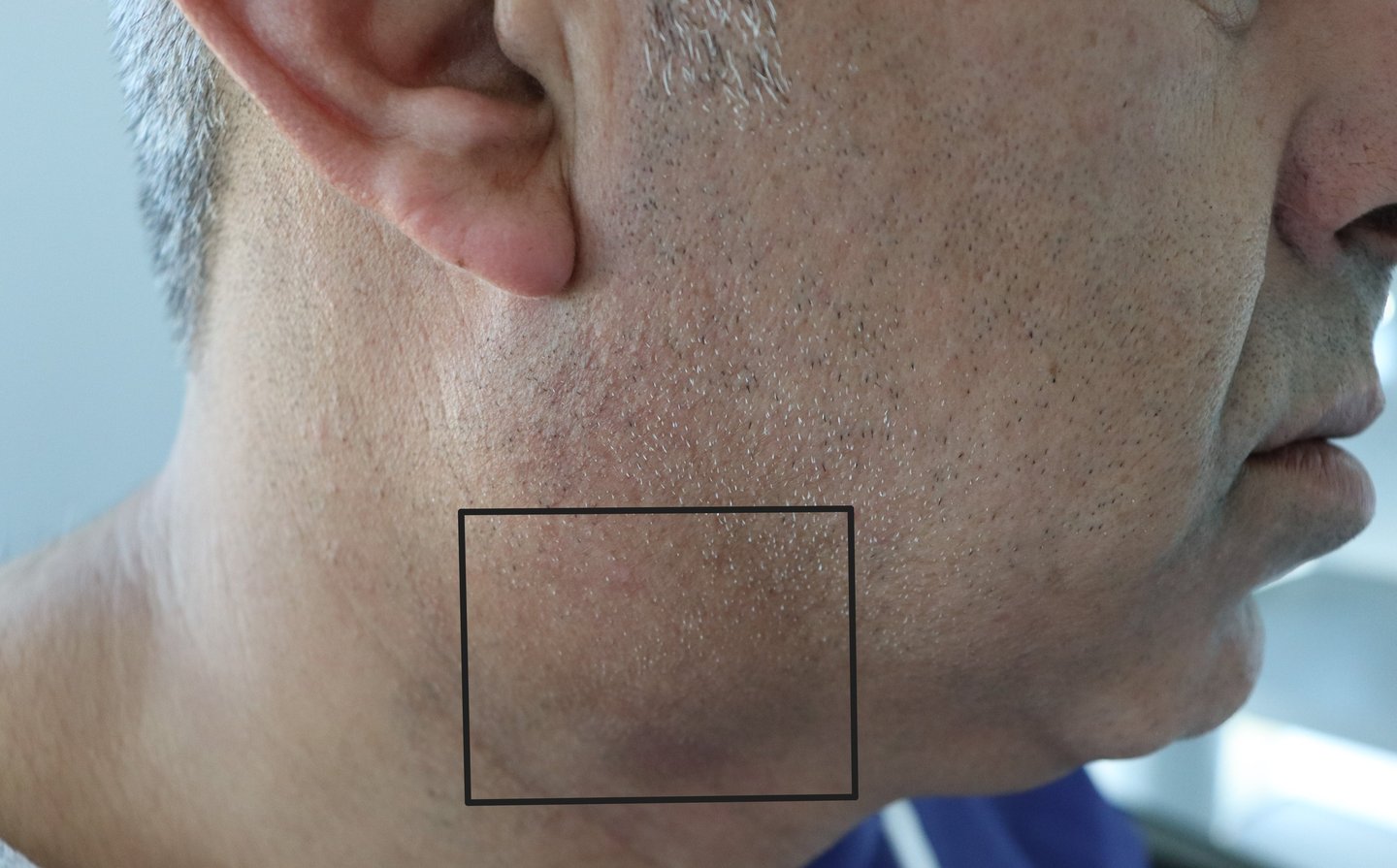
A 40-year-old male presented with a right submandibular mass that had been progressively enlarging over the past two months. The patient did not report any associated pain, fever, or difficulty swallowing.
History of Present Illness:
The patient noticed a growing mass on the right side of the neck, with no specific factors that aggravated or relieved the swelling. The mass was not associated with any discomfort, fever, or dysphagia.
Past Medical and Surgical History:
No significant medical or surgical history was reported.
Family History:
There was no relevant family history.
Examination:
General Examination:
Unremarkable.
Systemic Examination:
No abnormalities noted.
Neck Examination:
A right submandibular mass measuring approximately 4 cm in diameter was identified. The mass was fixed, non-tender, and appeared to be in the submandibular region.
Imaging Studies:
Neck Ultrasound Findings:
A well-defined, cystic mass was noted on the right side of the neck, located inferiorly to the low pole of the right parotid gland and partially covered by the anterior border of the upper part of the right sternocleidomastoid (SCM) muscle.
The mass was 49 × 33 × 28 mm in size, with a wall thickness of less than 3 mm. It appeared predominantly cystic, filled with internal echoes and debris, and showed hypovascularity. Fine internal septation was also noted. The surrounding tissue was compressed but otherwise normal. These findings were consistent with a complicated or infected second branchial cleft cyst.
CT Scan (with IV Contrast):
The scan revealed a well-defined cystic lesion anterior to the right sternocleidomastoid muscle, measuring 40 × 35 × 30 mm.
The lesion compressed the internal jugular vein and displaced the right submandibular gland. It showed a thick, enhancing wall with internal septation but no invasion into adjacent structures, suggesting an infected second branchial cleft cyst.
Fine Needle Aspiration Cytology (FNAC):
FNAC findings indicated features consistent with reactive lymphadenitis. No malignancy was identified. The recommendation was for total excision for definitive diagnosis.
Surgical Intervention:
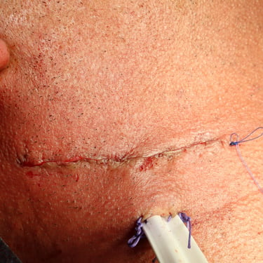
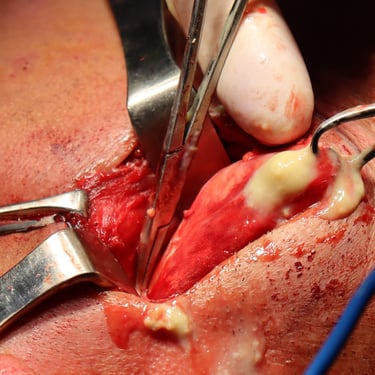
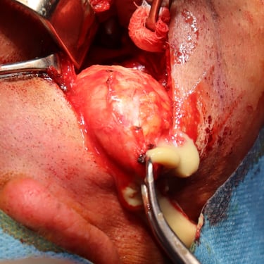
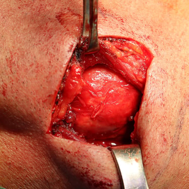
The patient underwent a total excision of the right submandibular mass.
Post-Operative Status:
The patient recovered well post-surgery with no complications and was discharged in good condition on the same day as the operation.
Histopathological Examination (HPE):
The histopathology report confirmed the diagnosis of a branchial cyst with no signs of malignancy.
Discussion:
This case presents a typical example of an infected second branchial cleft cyst, a congenital anomaly that results from incomplete obliteration of the second branchial cleft during embryogenesis. While these cysts are usually asymptomatic, infection or complications such as abscess formation can occur, presenting as an enlarging, tender mass.
Imaging studies like neck ultrasound and CT scans play an essential role in defining the cyst's location, size, and relationship with surrounding structures, and are crucial for preoperative planning. FNAC helps rule out malignancy and provides an initial diagnosis that aids in surgical management.
Surgical excision is the treatment of choice for branchial cleft cysts, and the Sistrunk operation, though typically used for thyroglossal duct cysts, is often adapted for cyst removal when appropriate. In this case, total excision of the cyst was performed, with no recurrence noted during follow-up.
Conclusion:
This case underscores the importance of accurate diagnosis and surgical management of congenital neck masses. Early intervention can prevent complications and recurrence, as demonstrated by the patient’s successful recovery. Regular follow-up is necessary to ensure there is no recurrence and to monitor for any potential complications related to adjacent structures.
Gallery
