Management of a 58-Year-Old Male with a Right Preauricular Mass
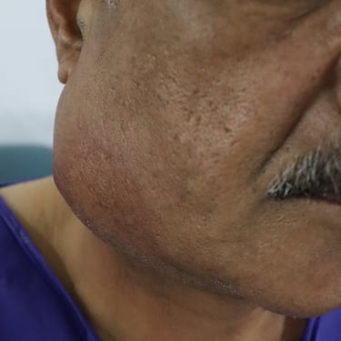
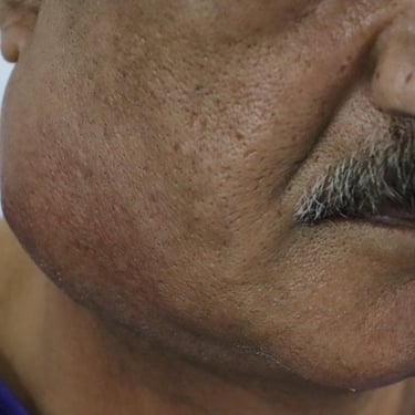
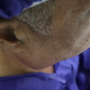
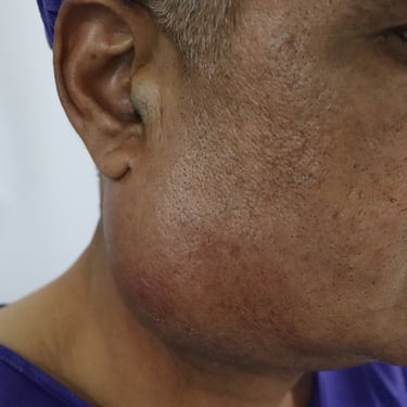
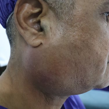
A 58-year-old male presented with a long standing Right preauricular mass for 2 years, developing pain and swelling in last week. His past medical, surgical, and drug history
SURGERY
8/6/20242 min read

Case Presentation:
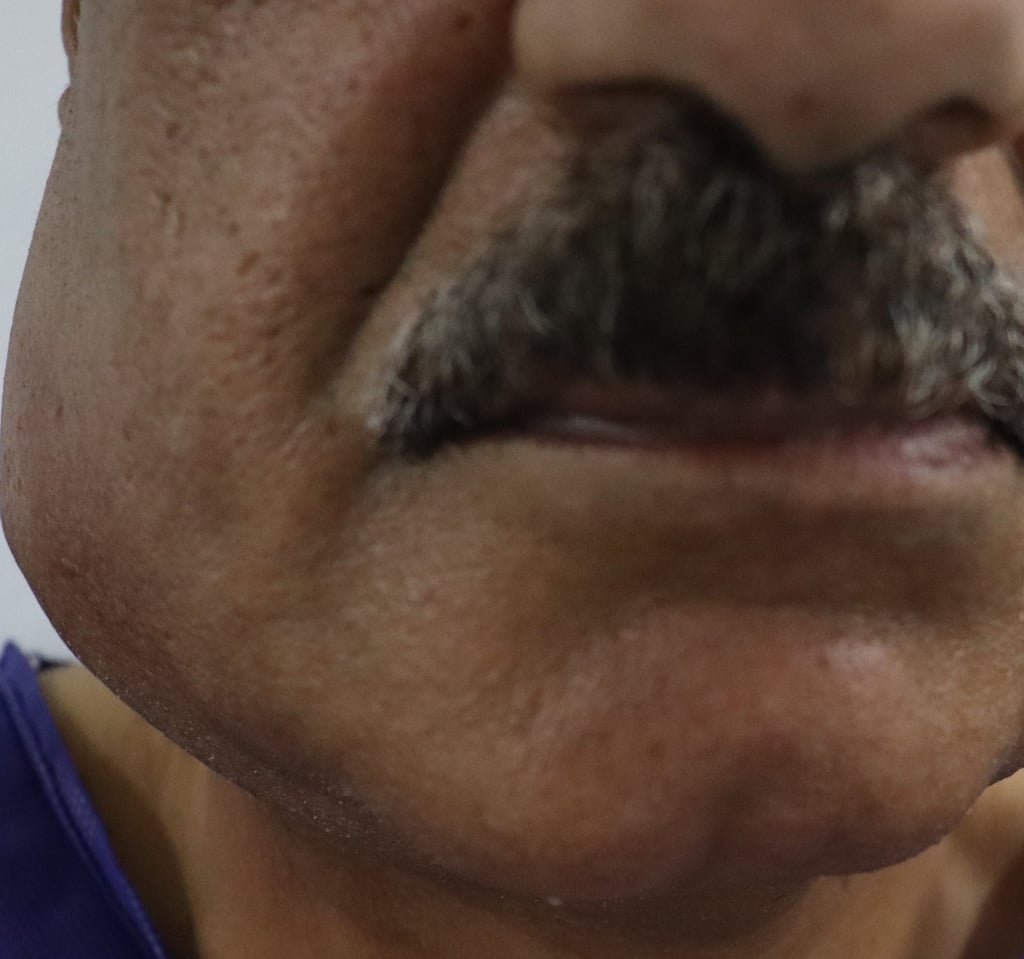
A 58-year-old male presented with a long-standing right preauricular mass that had been present for 2 years. Over the past week, the mass developed increased pain and swelling. His past medical, surgical, and drug history were unremarkable. The patient has been an active smoker for over 40 years.
Imaging Studies:
Neck Ultrasound Findings:
Right Parotid Gland: A large, complex, predominantly cystic mass (53 × 47 × 27 mm) was observed in the mid to lower third of the right parotid gland. The mass exhibited a 3 mm thick wall with mild peripheral vascularity and inhomogeneity. Edema was noted in the surrounding tissue and parenchyma of the gland, with ductal system dilatation, though no stones were visible. These findings were suggestive of a complicated or infected cystic mass, possibly hemorrhagic.
Lateral Cervical Lymphadenopathy (Right-sided): Multiple lymph nodes of variable sizes and shapes were seen, all well-defined with cortical thickness less than 5 mm. The largest lymph node, located in the right group II, measured 22 × 8.8 mm and was considered pathological. The nodes showed loss of hilar echotexture and mild vascularity, suggesting benign reactive changes.
Right Submandibular Gland: Normal in size but showed heterogeneous parenchymal echogenicity, without focal lesions or nodules.
Fine Needle Aspiration Cytology (FNAC):
FNAC from the right parotid gland mass revealed cystic content consistent with a pyogenic inflammatory process, suggesting an infected or hemorrhagic cyst.
FNAC from the right cervical pathological lymph node showed benign lymphoid cells, with no evidence of malignancy.
Surgical Intervention:
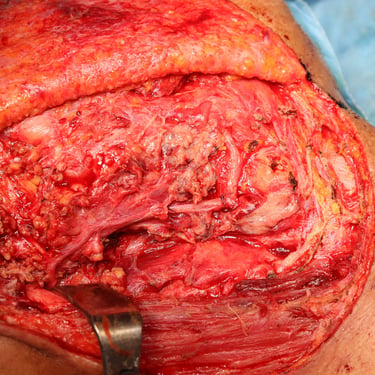
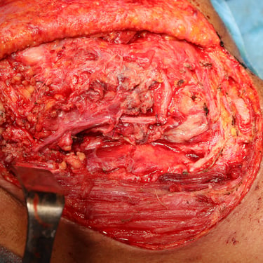
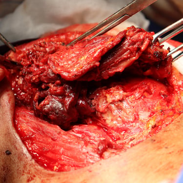
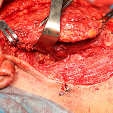
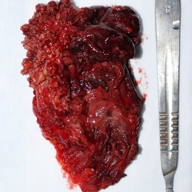
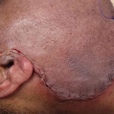
Given the imaging and cytological findings, a right superficial parotidectomy was performed. The decision was made to excise the parotid mass due to the possibility of infection or hemorrhage, as well as to rule out malignancy.
Histopathological Examination (HPE):
The histopathology report revealed a Warthin tumor with features of infarction, suppuration, and benign squamous metaplasia. Several benign reactive lymph nodes were also noted during the procedure, confirming the benign nature of the lymphadenopathy.
Discussion:
This case exemplifies the presentation and management of a Warthin tumor, a benign salivary gland tumor that is most commonly found in the parotid gland. The patient’s prolonged smoking history is a known risk factor for the development of Warthin tumors, which tend to present as slow-growing, painless masses, though infection or infarction may cause acute symptoms, such as pain and swelling, as observed in this patient.
Imaging, particularly ultrasound, plays a crucial role in evaluating the size, location, and characteristics of parotid masses. The presence of cystic components, vascularity, and surrounding edema raises suspicion for complications such as infection or hemorrhage. FNAC provides important cytological information to rule out malignancy and support the diagnosis.
Surgical excision, in the form of a superficial parotidectomy, is the treatment of choice for Warthin tumors, especially when complications like infarction or infection are present. The benign nature of the tumor is confirmed through histopathological analysis, which also revealed benign reactive lymph nodes consistent with the patient’s inflammatory response.
Conclusion:
This case highlights the importance of a thorough diagnostic workup, including imaging and FNAC, to manage parotid masses effectively. Warthin tumors, although benign, can present with acute symptoms due to complications, and surgical intervention is often necessary for definitive diagnosis and treatment. Smoking cessation and regular follow-up are recommended to monitor for recurrence or the development of new tumors.
Gallery

