Non-Hodgkin’s Lymphoma in an 85-Year-Old Male
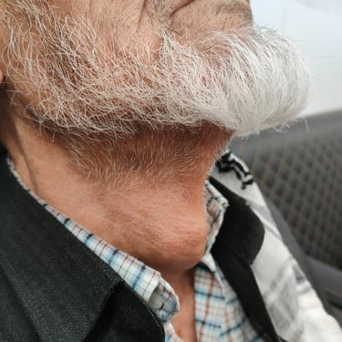
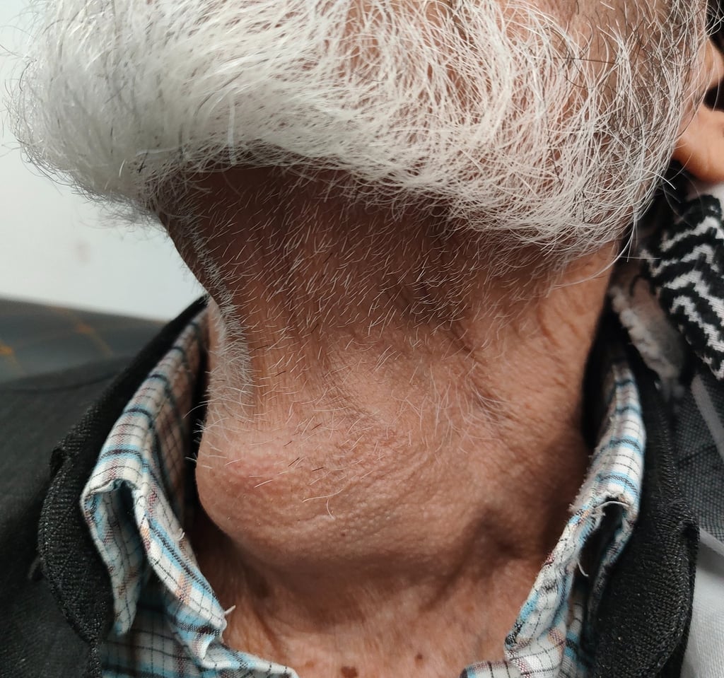
An 85-year-old male presented with a rapidly growing anterior neck mass, with no significant past medical or surgical history.
SURGERY
10/15/20231 min read

Case Presentation:
An 85-year-old male presented with a rapidly growing anterior neck mass, with no significant past medical or surgical history.
Neck CT Scan:
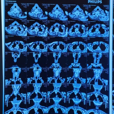
A neck CT scan revealed an anterior neck lesion measuring approximately 5.4 × 4 × 3 cm, located in relation to the thyroid gland. The mass extended from the isthmus and reached below the hyoid bone. It had a non-enhancing cystic component at the upper aspect, without calcification, and showed signs of tracheal compression and retrosternal extension.
Neck Ultrasound:
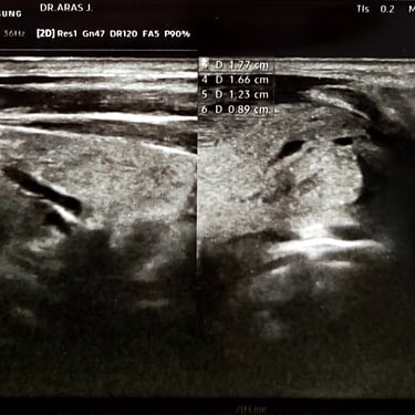
Ultrasound findings revealed a few bilateral small TR3 nodules, along with a large TR4 nodule measuring 47 × 46 × 18 mm. The nodule was well-defined, with a regular surface, solid, hypoechoic, and mildly vascular. The ultrasound also showed bilateral small inflammatory cervical lymph nodes. Fine Needle Aspiration (FNA) was suggested.
Laboratory Tests:
The laboratory tests revealed high CRP levels, and the TSH was 1.13 (Normal).
FNAC:
The Fine Needle Aspiration Cytology (FNAC) was suggestive of Non-Hodgkin’s lymphoma involving the thyroid gland, and a tissue biopsy was recommended for confirmation.
Core Biopsy:
The core biopsy results confirmed Non-Hodgkin’s lymphoma, with features suggestive of diffuse large B-cell lymphoma. Immune stains were recommended for further evaluation.
Immune Stains:
The immune stains were as follows:
CD3, CD10, CD20, CD23, CD45, CD79, MUM1, BCL2, BCL6 were all positive
Cyclin D1 was negative.
These reactions, in conjunction with the cytological features, were consistent with a diagnosis of diffuse large B-cell lymphoma.
Image Gallery
