Papillary Thyroid Carcinoma in a Pregnant Patient with Long-Standing Graves’ Disease
A 38-year-old female, previously diagnosed with Graves’ disease and on long-term anti-thyroid medication, presented for routine thyroid follow-up during her 15th week of pregnancy. She sought medical attention for dosage regulation of her anti-thyroid medication. On clinical examination, palpable thyroid nodules were noted, prompting further laboratory and imaging evaluations.
SURGERYHEAD AND NECKVIDEO
5/1/20251 min read

Clinical Presentation:
A 38-year-old female, previously diagnosed with Graves’ disease and on long-term anti-thyroid medication, presented for routine thyroid follow-up during her 15th week of pregnancy. She sought medical attention for dosage regulation of her anti-thyroid medication. On clinical examination, palpable thyroid nodules were noted, prompting further laboratory and imaging evaluations.
Laboratory and Imaging Findings:
Laboratory tests showed a TSH level of 1.72 mIU/L and FT4 of 12.7 pmol/L, indicating stable thyroid function. A previous TSH receptor antibody (TRAb) test was initially positive at 8.84 IU/L but had become negative (1.17 IU/L) on the latest assessment, suggesting disease modulation.
Thyroid ultrasound revealed a heterogeneous gland with multiple nodules:
A solid hypoechoic nodule (10×8×5 mm) at the right lobe-isthmus junction, with microcalcifications and increased perinodular-intranodular vascularity (TR5).
A similar TR5 nodule (9×8×7 mm) in the isthmus.
Multiple lymph nodes (LNs) around the gland, with the largest (17×11 mm) located at the suprasternal notch.
Bilateral lateral cervical lymph nodes, the largest measuring 11×3 mm near the left upper pole, with preserved hilum and no suspicious sonographic features.
Fine Needle Aspiration (FNA) and Diagnosis
Given the suspicious TR5 nodules, FNA was performed on the right thyroid lobe-isthmus nodule. Cytology confirmed a Bethesda VI lesion, consistent with papillary thyroid carcinoma (PTC).
Preoperative Assessment and Surgical Plan:
The patient underwent a comprehensive preoperative evaluation before surgery. Laboratory results showed a thyroglobulin (Tg) level of 46.5 ng/mL and a serum calcium level of 9.43 mg/dL, indicating no immediate concerns regarding thyroid function or calcium balance. Viral screening was negative, and vocal cord assessment confirmed normal movement, ensuring no pre-existing vocal cord dysfunction.
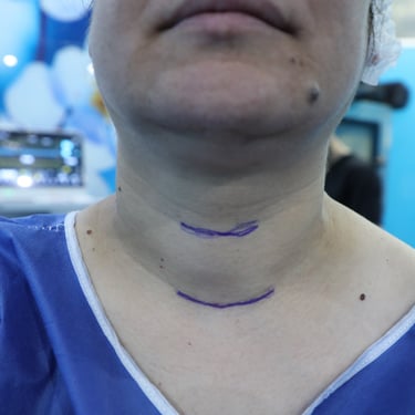
The patient was scheduled for total thyroidectomy with bilateral central lymph node dissection (CLND).
Surgical Outcome and Histopathological Findings:
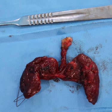
The patient underwent total thyroidectomy with bilateral CLND. Histopathological examination confirmed papillary thyroid carcinoma (PTC) in the background of Graves’ disease. She was scheduled for post-surgical follow-up and further oncologic evaluation.
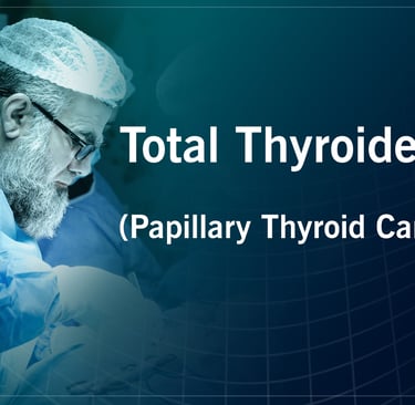
Head and Neck Surgeon: Prof. Dr. Abdulwahid Mohammed Salih
Co-surgeon: Dr. Yadgar Abdulhameed Saeed
Assistant: Dr. Harun Amanj Ahmed & Dr. Dindar Hussein Hama

