Parathyroid Cyst in a 46-Year-Old Female
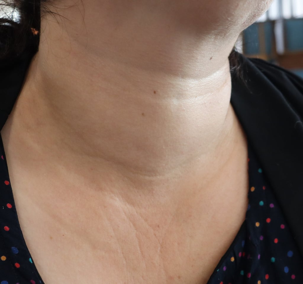
A 46-year-old female presented with a one-week history of dysphagia, generalized body weakness, and associated sweating. There were no additional symptoms, and the patient had no significant past medical or surgical history. On physical examination, there were no remarkable findings.
SURGERY
10/1/20231 min read

Case Presentation:
A 46-year-old female presented with a one-week history of dysphagia, generalized body weakness, and associated sweating. There were no additional symptoms, and the patient had no significant past medical or surgical history. On physical examination, there were no remarkable findings.
Blood Investigations:
Laboratory tests revealed thyroid function within normal limits (TSH: 2.46, FT4: 14, ATPO: 16.4), but parathyroid hormone (PTH) levels were elevated (112.8), along with a serum calcium level of 9.54.
Imaging Studies:
Neck ultrasonography identified a well-defined, thin-walled cystic nodule measuring 46 × 30 × 25 mm located posterior to the thyroid, extending retrosternally and crossing the midline. A subsequent CT scan of the neck described the lesion as a 47 × 43 × 31 mm fluid-density thin-walled cystic structure in the superior mediastinum, initially suggestive of an esophageal duplication cyst, with a differential diagnosis that included a bronchogenic cyst.
Endoscopic Evaluation:
Esophagogastroduodenoscopy (OGD) revealed normal mucosa with no signs of compression or features indicative of an esophageal duplication cyst.
Multidisciplinary Team Discussion:
This challenging case was extensively discussed within a Multidisciplinary Team (MDT) due to the atypical presentation and diagnostic uncertainties. The team collectively decided on an excision procedure, highlighting the importance of collaborative, team-based decision-making in managing complex cases.
Surgical Intervention:
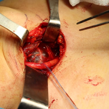
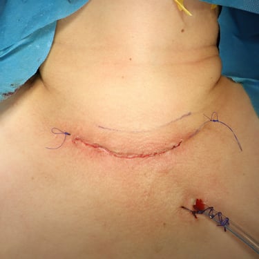
A trans-cervical approach was employed for cyst excision, revealing a large, thin-walled cyst with clear fluid content during gross examination.
Postoperative Course:
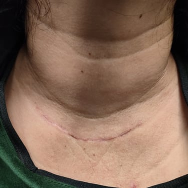
Postoperative investigations revealed a decrease in PTH levels to 62, and calcium levels were within the normal range (9.14). The patient experienced symptomatic improvement postoperatively.
Histopathological Analysis:
The excised specimen revealed multiple rubbery ruptured cystic pieces, with the largest measuring 2.5 × 1 cm. The final diagnosis was a parathyroid cyst, with no evidence of malignancy. The cystic wall showed simple cuboidal cells and contained mature parathyroid tissue along with adipocytes.
Image Gallery

