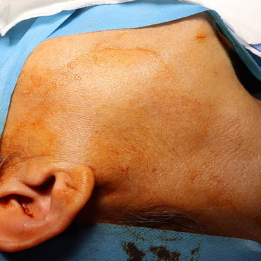
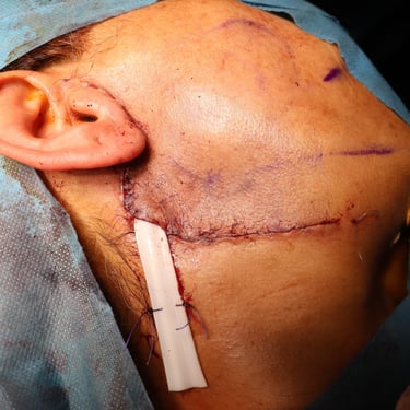
Pleomorphic Adenoma of the Right Parotid Gland
A 51-year-old female presented with a right parotid swelling of 1-year duration. The swelling was asymptomatic with no associated pain, discharge, or functional impairment.
SURGERYHEAD AND NECK
1/30/20251 min read

Case Presentation:
A 51-year-old female presented with a right parotid swelling of 1-year duration. The swelling was asymptomatic with no associated pain, discharge, or functional impairment.
Past Medical History (PMH): Negative
Past Surgical History (PSH): Negative
Medication History: Negative
Investigations:
None
Ultrasound (US):
No significant cervical lymphadenopathy.
Normal parotid and submandibular glands, with no focal lesions.
A small, complex nodule measuring 16 x 14 x 11 mm was identified in the superficial lobe of the right parotid gland. The nodule had increased in size compared to a previous ultrasound, raising suspicion for a neoplasm.
Fine Needle Aspiration Cytology (FNAC):
Diagnosis: Neoplasm, benign (Milan System Category IVA).
Cytological Suggestion: Pleomorphic adenoma (benign mixed salivary gland tumor).
Surgery:
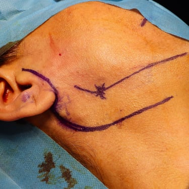
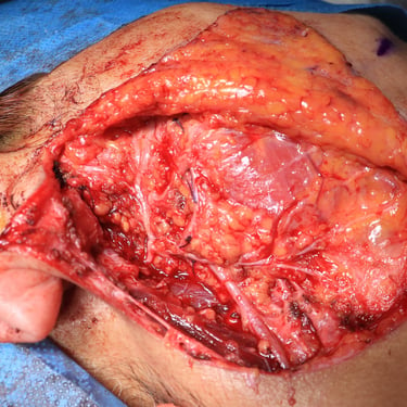
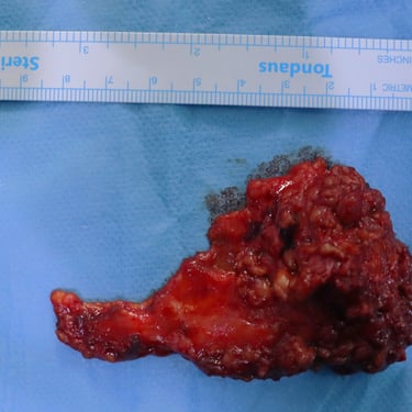
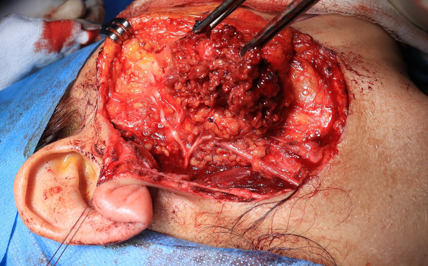
The patient underwent a superficial parotidectomy of the right parotid gland.
Histopathological Examination (HPE):
Final Diagnosis: Pleomorphic Adenoma
Composed of a mixture of epithelial and stromal elements.
The tumor was well-encapsulated with no evidence of malignant transformation.
Margins were clear of the lesion.
Postoperative Course:
The patient recovered well with no complications, such as facial nerve injury or hematoma.
Follow-up ultrasound and clinical examination showed no recurrence or residual disease.
Discussion:
Pleomorphic adenoma, also known as benign mixed tumor, is the most common benign salivary gland neoplasm, with a predilection for the parotid gland. Although generally benign, pleomorphic adenomas have the potential for malignant transformation into carcinoma ex pleomorphic adenoma, especially if left untreated for long periods.
