Post-Thyroidectomy Sinus Formation in a 34-Year-Old Female
A 34-year-old female presented with a chronic anterior neck lesion that had been present for 20 years following a thyroidectomy. The patient had no significant past medical history and was on Thyroxine (50µg) once daily.
SURGERY
11/1/20231 min read

Case Presentation:
A 34-year-old female presented with a chronic anterior neck lesion that had been present for 20 years following a thyroidectomy. The patient had no significant past medical history and was on Thyroxine (50µg) once daily.
Clinical Findings:
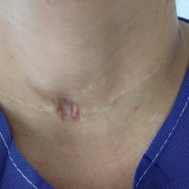
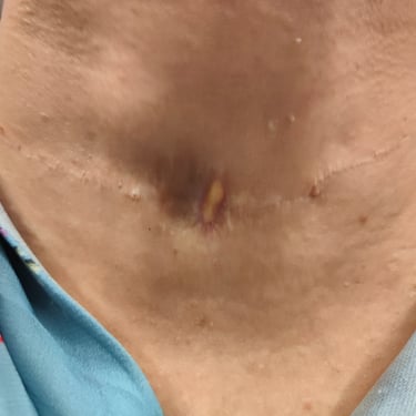
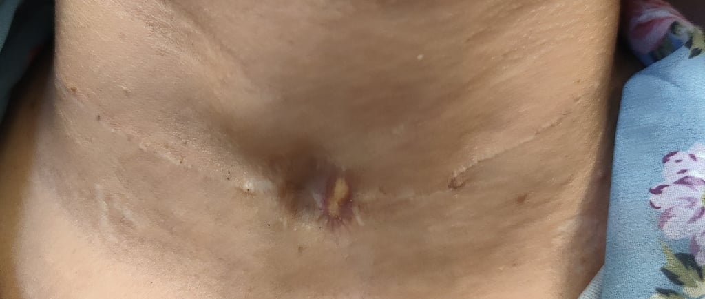
Upon examination, a brown circular lesion was observed on the anterior neck with evidence of discharge. There were no signs of inflammation. The lesion was located over a collar scar from her previous thyroidectomy.
Laboratory Findings:
Thyroid function tests were within normal limits, as indicated by the following results:
TSH: 3.75 (Normal)
Free T4: 16.37 (Normal)
ATPO: 13.73 (Normal)
Imaging Findings:
Neck ultrasound revealed remnant thyroid tissue in both lobes, with the right lobe measuring 27 × 19 × 19 mm and the left lobe 45 × 14 × 11 mm. Both remnants showed heterogeneous texture, with small nodules observed in both lobes. The largest nodule in the right lobe was 16 × 9 × 7 mm TR2, and the largest in the left lobe was
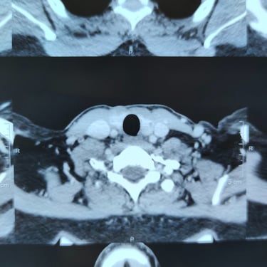
7 × 4 × 5 mm TR3. No highly suspicious lesions were identified, and there was a clear operative bed in the isthmus. No significant cervical lymphadenopathy was noted. A CT scan further confirmed bilateral remnant thyroid tissue and showed a focal dimpling of the anterior neck skin with subcutaneous fat loss, but no collection, sinus, or fistula tract was seen.
Therapeutic Management:
The patient was scheduled for redo thyroidectomy. Under general anesthesia, the procedure was carried out in the supine position. Both recurrent laryngeal nerves (RLN) were explored and preserved. The incision was made superior and inferior to the lesion, and both the lesion and the old scar were excised.
Image Gallery
