
Recurrent Moderately Differentiated Squamous Cell Carcinoma of the Lower Lip in a 58-Year-Old Male
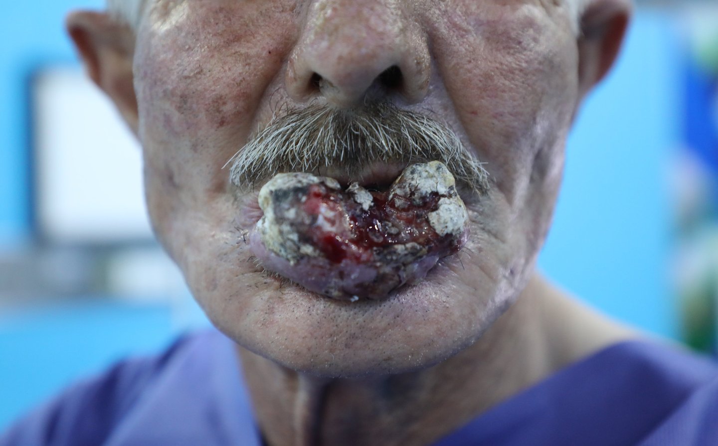
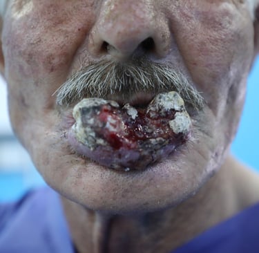
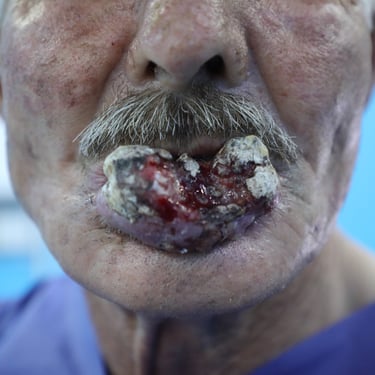
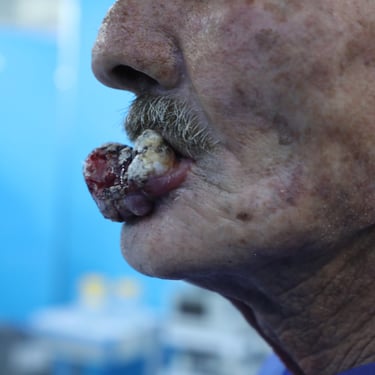
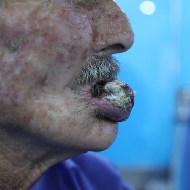
A 58-year-old male presented to the surgical oncology clinic with a progressively enlarging ulcerative lesion on the lower lip, present for approximately one year. He is a known case of hypertension, for which he is receiving antihypertensive medications. The patient has a longstanding history of heavy tobacco use and remains an active smoker. He does not have a known history of diabetes, hepatitis, or other significant comorbidities. The persistent nature and progressive enlargement of the lesion prompted further investigation for malignancy.
SURGERYHEAD AND NECK
7/22/20253 min read
Patient Information:
A 58-year-old male presented to the surgical oncology clinic with a progressively enlarging ulcerative lesion on the lower lip, present for approximately one year. He is a known case of hypertension, for which he is receiving antihypertensive medications. The patient has a longstanding history of heavy tobacco use and remains an active smoker. He does not have a known history of diabetes, hepatitis, or other significant comorbidities. The persistent nature and progressive enlargement of the lesion prompted further investigation for malignancy.
Ultrasound Findings:
Neck ultrasonography revealed a large, full-thickness, solid, hypoechoic, and hypervascular ulcerating mass arising from the lower lip, measuring approximately 41 × 21 × 19 mm. The lesion showed irregular margins and sonographic features of tissue invasion, raising strong suspicion for an aggressive malignancy. Multiple bilateral lymph nodes were visualized, predominantly on the left side. These included a 16 × 14 mm lymph node in the left submandibular region, a 10 × 6 mm node in the submental group, and a 5 × 3 mm node in the left parotid group. The lymph nodes were solid, heterogeneously hypoechoic, and demonstrated features consistent with pathological involvement.
Staging Imaging:
A contrast-enhanced CT scan of the chest, abdomen, and pelvis was performed to evaluate for possible distant metastases. The scan showed mild bilateral centrilobular emphysematous changes and linear fibrotic nodules at the apical segments of both upper lobes, suggesting chronic pulmonary changes likely secondary to smoking. However, there were no suspicious pulmonary nodules, no pathological mediastinal or hilar lymphadenopathy, and no signs of metastatic disease. The abdominal and pelvic organs, including the liver, kidneys, pancreas, spleen, gastrointestinal tract, and adrenal glands, were all unremarkable. There was no evidence of lymphadenopathy or mass lesions in the retroperitoneal or pelvic regions. Bone windows revealed diffusely decreased bone density but no obvious lesions suggestive of metastasis. Overall, no evidence of distant metastasis was found.
Cytology and Laboratory Investigations:
Fine-needle aspiration cytology (FNAC) was performed on a left group I cervical lymph node. Cytologic analysis revealed benign lymphoid cells with no evidence of malignancy. Routine blood investigations were largely unremarkable, with serum creatinine at 0.778 mg/dL and blood urea slightly elevated at 46.0 mg/dL. Fasting glucose was mildly elevated at 116 mg/dL, which may warrant follow-up but did not affect immediate surgical planning. Viral screening for hepatitis B, hepatitis C, and HIV was negative.
Preoperative Evaluation and Surgical Plan:
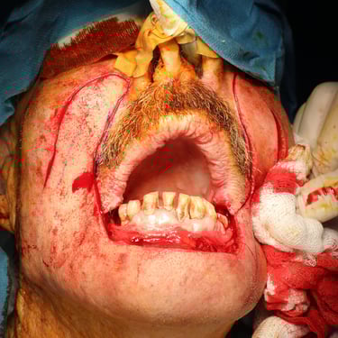
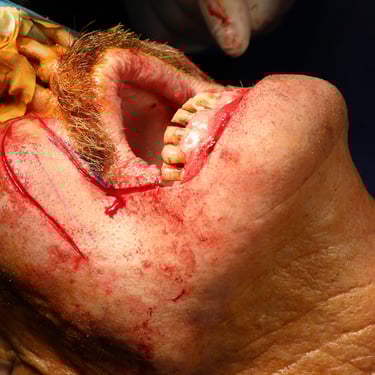
In light of the clinical and radiologic evidence, the patient was scheduled for definitive surgical management. The operative plan included a wide local excision of the lower lip lesion, bilateral submandibular gland excision, and selective neck dissection involving level III lymph nodes. Intraoperative labeling was used to mark the excised tissues, including identification of the left-sided lymph nodes and submandibular gland with sutures. The patient was considered a surgical candidate despite his chronic smoking history, and his overall cardiovascular and renal function was deemed adequate for general anesthesia.
Surgical Intervention:
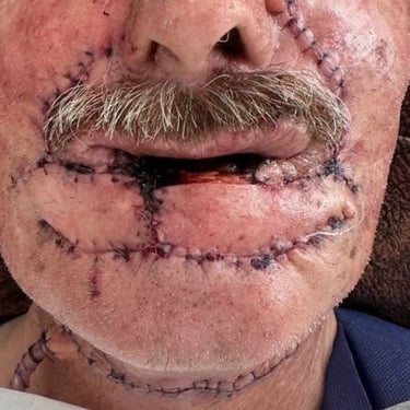
The patient underwent wide local excision of the lower lip lesion along with bilateral submandibular gland removal and level III cervical lymph node dissection. The surgical procedure was completed without complications, and all excised specimens were submitted for detailed histopathological evaluation.
Histopathological Examination (HPE):
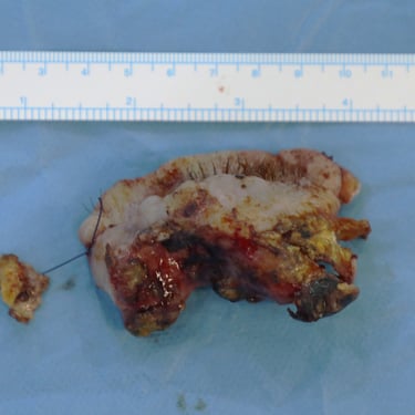
Microscopic analysis confirmed squamous cell carcinoma of the lower lip, moderately differentiated (Grade 2). The tumor measured 4.5 cm in maximal dimension with a thickness of 2.5 cm and depth of invasion of 1.6 cm. The lesion invaded through subcutaneous fat and into the skeletal muscle of the lip, and ulceration was present on the surface. Lymphovascular invasion was identified, indicating increased risk of dissemination, but no perineural invasion was seen. All surgical margins were negative, with the closest being 0.9 cm (left and inferior margins). A total of 26 regional lymph nodes were examined, and none contained metastatic carcinoma. Both the right and left submandibular glands were histologically unremarkable. Additional findings included solar elastosis, consistent with chronic sun exposure. The final pathologic staging was pT3 pN0, indicating locally advanced tumor without regional nodal metastasis.
Postoperative Management and Follow-Up Plan:
Given the tumor’s size, depth of invasion, and the presence of lymphovascular invasion—despite clear margins and absence of nodal involvement—the patient is considered at intermediate to high risk for recurrence. A multidisciplinary tumor board evaluation is recommended to determine the need for postoperative radiotherapy to reduce local recurrence risk. Smoking cessation counseling is a critical component of his postoperative care plan due to the significant contribution of tobacco use to recurrence and secondary malignancy risk. Surveillance will include regular physical examinations, neck imaging (preferably ultrasound or MRI), and periodic chest imaging, particularly in light of his underlying pulmonary changes and smoking history. Long-term follow-up will focus on early detection of local recurrence, nodal spread, or distant metastases.
Gallery
Address
Majid Bag Main Street, Beside University of Sulaymaniyah Old Campus, Madam Mitterrand, Sulaymaniyah, Iraq
Contacts
info@ssthyroid.com