Recurrent Multinodular Goiter in a Patient with Prior Thyroid Surgery
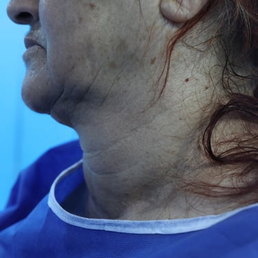
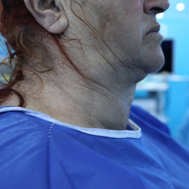
A 70-year-old male presented with a long-standing anterior neck swelling. On physical examination, the swelling was classified as grade III, large in size, firm, slightly mobile, and had a soft surface. The patient denied any significant symptoms such as dyspnea, dysphagia, or voice changes.
SURGERYHEAD AND NECKVIDEO
5/17/20252 min read

Patient Presentation:
A 70-year-old male presented with a long-standing anterior neck swelling. On physical examination, the swelling was classified as grade III, large in size, firm, slightly mobile, and had a soft surface. The patient denied any significant symptoms such as dyspnea, dysphagia, or voice changes.
Medical and Surgical History:
The patient had no notable past medical history and was not on any medications. However, his surgical history was significant for a thyroid operation performed approximately 35 years ago, which was likely a subtotal thyroidectomy. There was no relevant family history of thyroid disease.
Laboratory Investigations:
To evaluate his thyroid status and rule out complications from the previous surgery, laboratory tests were performed. Results showed a TSH of 0.352 uIU/mL and FT4 of 15.6 pmol/L, indicating a euthyroid state. Serum calcium was within normal limits at 10.1 mg/dL. PTH was elevated at 71.0 pg/mL, suggesting mild secondary hyperparathyroidism or post-surgical compensatory changes. Calcitonin was 0.518 pg/mL, and thyroglobulin was significantly elevated at 153 ng/mL, likely due to the presence of multiple nodules.
Neck Ultrasound:
Ultrasound of the thyroid demonstrated enlargement of both lobes and the isthmus, with multiple bilateral nodules of varying size and character. Most nodules were echoic and partially cystic. The largest was located in the left lobe near the midline, measuring 21 × 14 mm, with internal vascularity and a TR3 classification, indicating intermediate suspicion.
CT Neck and Chest:
CT imaging revealed a multinodular goiter with grade III retrosternal extension and significant compressive effects, including mild rightward tracheal deviation and marked tracheal narrowing, with only a slit-like airway remaining. These findings raised concern for progressive compressive symptoms despite the patient’s lack of acute complaints.
Vocal Cord Assessment:
Flexible laryngoscopy was performed and showed normal bilateral vocal cord mobility, with no evidence of paralysis or previous surgical injury.
Surgical Management:
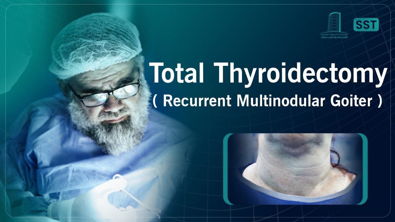
Given the findings of recurrent goiter with retrosternal extension and tracheal compression, the patient underwent a total thyroidectomy. The surgery was performed under general anesthesia using a collar incision, with the patient in a supine, extended neck position. The operation was completed in approximately 1.5 hours with no intraoperative complications.
Histopathological Examination:
The final histopathological diagnosis was consistent with benign follicular nodular disease with secondary changes. No malignancy was identified in the thyroid tissue. These findings confirmed a diagnosis of recurrent multinodular goiter.
Head and Neck Surgeon: Dr Abdulwahid M. Salih
Co-Surgeon: Dr. Yadgar Abdulhameed Saeed & Dr. Karzan Mohammed Salih Hassan
Assisstant: Dr. Harun Amanj Ahmed

