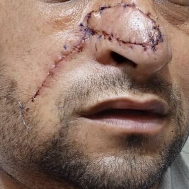
Right Nasal Squamous Cell Carcinoma with Postoperative Surveillance and Supra-omohyoid Neck Dissection
A 36-year-old male presented for follow-up one month after excision of a right nasal lesion diagnosed as squamous cell carcinoma (SCC). He is an active smoker and has no notable past medical history. His surgical history includes an ear surgery under general anesthesia three years prior and a recent excision of the right nasal lesion. Given the oncologic nature of the excised lesion, further evaluation was pursued to assess potential locoregional spread. The patient was referred to dermatology and underwent ultrasonography of the head and neck.
SURGERYHEAD AND NECK
6/17/20252 min read

Clinical Presentation:
A 36-year-old male presented for follow-up one month after excision of a right nasal lesion diagnosed as squamous cell carcinoma (SCC). He is an active smoker and has no notable past medical history. His surgical history includes an ear surgery under general anesthesia three years prior and a recent excision of the right nasal lesion. Given the oncologic nature of the excised lesion, further evaluation was pursued to assess potential locoregional spread. The patient was referred to dermatology and underwent ultrasonography of the head and neck.
Laboratory and Imaging Investigations:
Ultrasound of the neck demonstrated postoperative changes at the right nasal site with an ill-defined, heterogeneously hypoechoic focus suggestive of residual or reactive changes. Additionally, multiple bilateral cervical lymph nodes, predominantly on the right (levels I and II), were noted. Most lymph nodes were well-defined with cortical thickness less than 3 mm and preserved normal shape and hilar echotexture. However, one right submandibular lymph node measured 14 × 6.7 mm and exhibited loss of the fatty hilum, rendering it suspicious for malignancy on imaging. Thyroid, parotid, and submandibular glands were normal in size and echotexture, with no focal lesions identified.
Cytology and Dermatologic Evaluation:
Ultrasound-guided fine-needle aspiration (FNA) of the suspicious right submandibular lymph node was performed. Cytology revealed benign lymphoid cells with no evidence of malignancy. Dermatology consultation recommended either a repeat incisional biopsy or a review of the previously excised pathology slides, as the initial histopathological assessment was not performed at the current center. Slide review confirmed a diagnosis of keratoacanthoma-like squamous cell carcinoma, well-differentiated (Grade 1), with a tumor size of 1.4 cm, a depth of invasion of 0.4 cm, and a thickness of 0.6 cm. The tumor extended into the subcutaneous fat and superficial skeletal muscle bundles, with ulceration present. No lymphovascular or perineural invasion was noted. Margins were close, with the tumor touching the deep resection margin and being less than 0.1 cm from the nearest peripheral margin. The tumor was staged pathologically as pT1, and background solar elastosis was identified.
Multidisciplinary Team (MDT) Discussion and Surgical Management:
The case was discussed in a multidisciplinary tumor board. Given the presence of close surgical margins, dermal invasion, and potential for microscopic spread, the MDT recommended revision surgery. A wide local excision of the right nasal skin was planned in conjunction with a supraomohyoid neck dissection encompassing cervical lymph node groups I through IV.
Surgical Procedure:
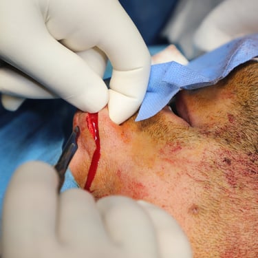
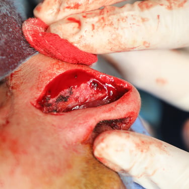
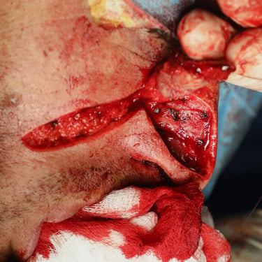
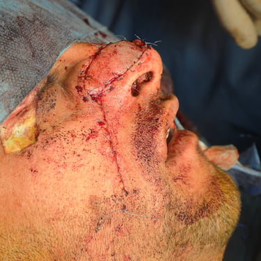
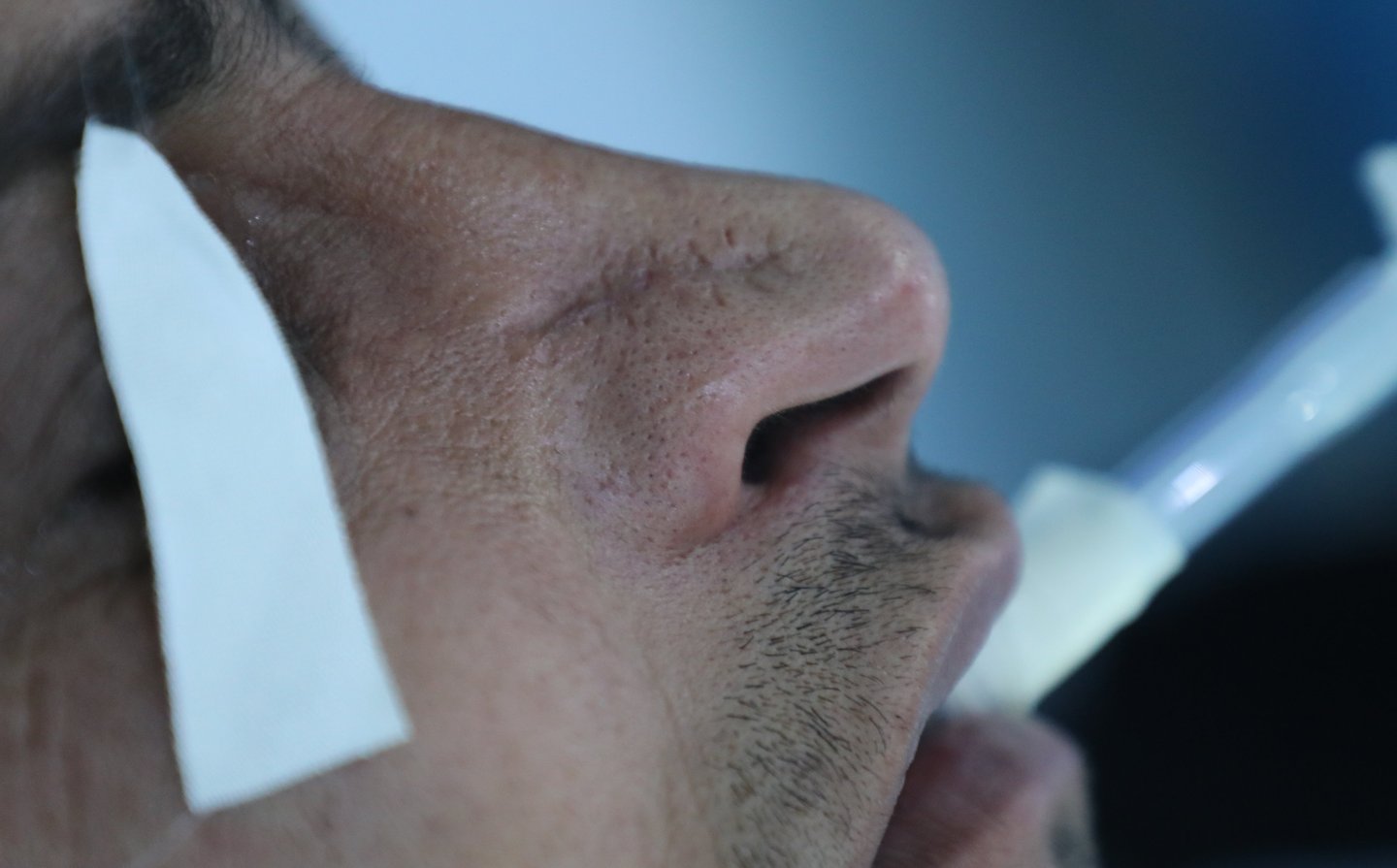
The patient underwent a right nasal skin wide local excision along with supraomohyoid neck dissection (levels I–IV). The surgery proceeded without intraoperative complications, and tissue specimens were submitted for histopathological evaluation.
Histopathology Results:
Histopathological examination of the wide local excision revealed dermal fibrosis with a giant cell reaction to retained suture material. There was moderate mononuclear inflammatory infiltrate, a small inclusion cyst, and features of solar elastosis. Importantly, there was no evidence of residual carcinoma or dysplasia in the re-excised skin. The supraomohyoid neck dissection yielded 25 lymph nodes, all of which were negative for tumor involvement, confirming the absence of regional metastasis.
Gallery

