Right-Sided Cervical Lymphangioma in a 35-Year-Old Female: Excision and Benign Outcome
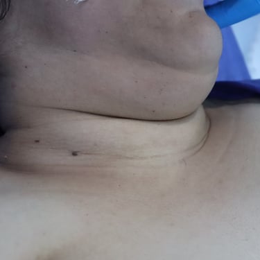
A 35-year-old female presented with an anterior neck swelling that had been gradually enlarging over the past one year. She reported no associated symptoms such as pain, dysphagia, or dyspnea. Her past medical and surgical history was unremarkable, and there was no family history of neck masses or endocrine disorders. On physical examination, the neck mass was soft to mildly firm in consistency and non-tender. There were no overlying skin changes, and no signs of local invasion were noted.
SURGERYHEAD AND NECK
5/6/20251 min read

Patient Presentation:
A 35-year-old female presented with an anterior neck swelling that had been gradually enlarging over the past one year. She reported no associated symptoms such as pain, dysphagia, or dyspnea. Her past medical and surgical history was unremarkable, and there was no family history of neck masses or endocrine disorders. On physical examination, the neck mass was soft to mildly firm in consistency and non-tender. There were no overlying skin changes, and no signs of local invasion were noted.
Laboratory and Imaging Evaluation:
Initial laboratory evaluation confirmed that the patient was euthyroid, with no biochemical abnormalities. Neck ultrasound revealed a large, well-defined lobulated cystic mass measuring approximately 22 × 11 × 9 cm, originating from the right side of the neck, beginning under the right sternocleidomastoid muscle and extending from the mastoid process to the retroclavicular region, with partial involvement of the posterior triangle. The lesion was predominantly cystic with mild vascularity, causing mild compression of surrounding structures but without signs of tissue invasion. The sonographic impression was most consistent with a lymphangioma.
The thyroid gland appeared normal in size, echotexture, and vascularity, with no focal lesions. Additionally, the parotid and submandibular glands were normal, and there was no significant cervical lymphadenopathy.
Multidisciplinary Discussion and Surgical Intervention:
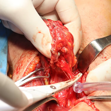
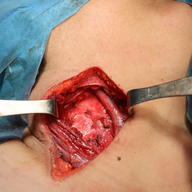
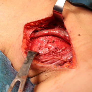
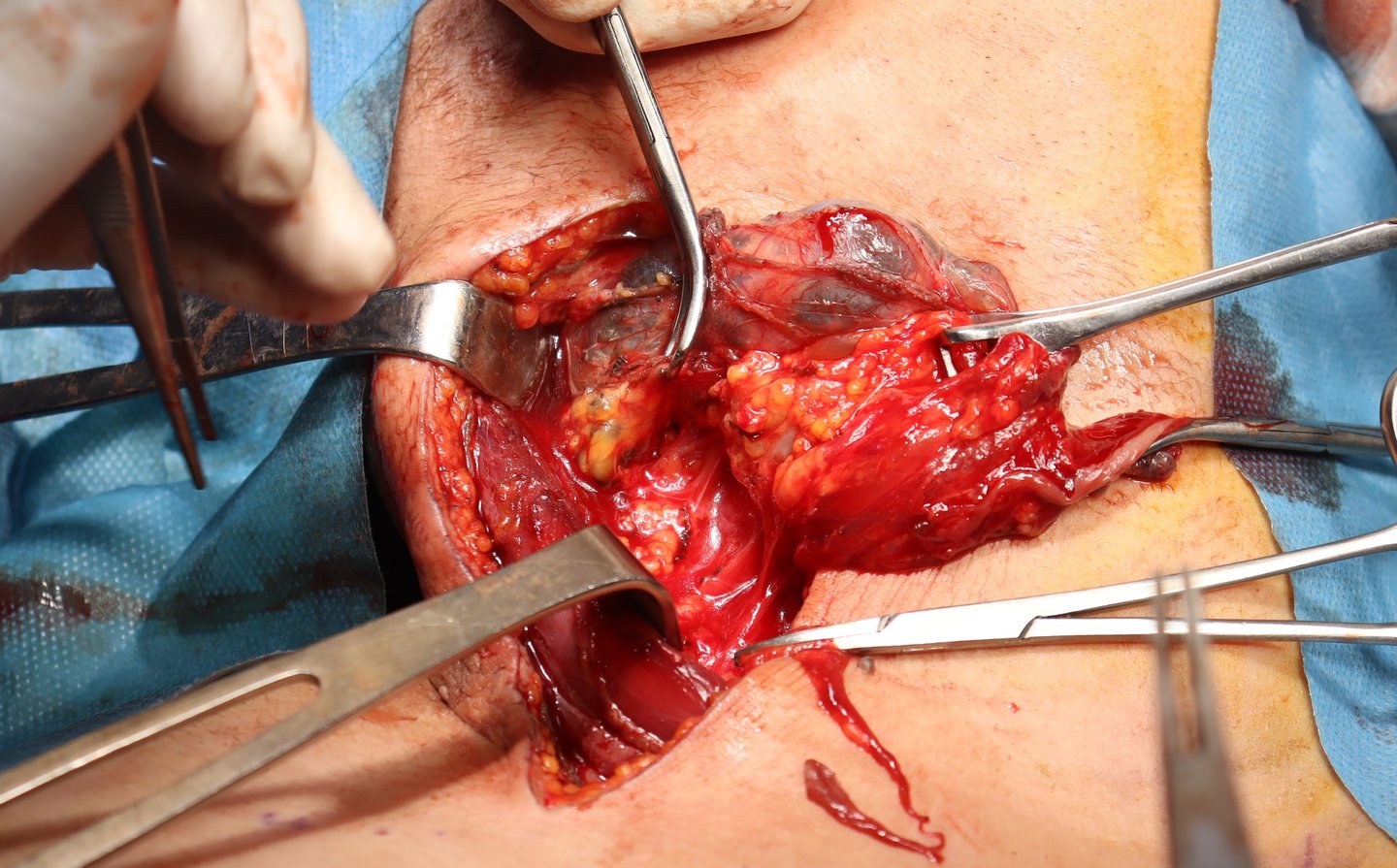
Following discussion at the multidisciplinary tumor board (MDT), the decision was made to proceed with surgical excision of the mass due to its size, progressive enlargement, and compressive effects. The patient underwent right-sided neck lymphangioma excision under general anesthesia. The procedure was uneventful, and all visible cystic tissue was completely removed.
Histopathology Report:
Histological examination of the resected specimen confirmed the diagnosis of a lymphangioma, with associated benign reactive lymph nodes. There was no evidence of malignancy, supporting the clinical and radiological impression of a benign congenital lymphatic lesion.
Postoperative Outcome and Follow-Up:
The patient recovered well postoperatively without complications. At follow-up visits, she remained in good condition, with no signs of recurrence or residual swelling. Surveillance imaging was planned as part of routine monitoring, although recurrence risk was considered low after complete excision.
Gallery
