Thyroid Imaging, Reporting and Data System (TIRADS)
Ultrasound features: Scoring is determined five categories. The higher the cumulative score, the higher the TR and the high the likelihood of malignancy.
RADIOLOGY
1/1/20233 min read

Ultrasound features
Scoring is determined five categories. The higher the cumulative score, the higher the TR and the high the likelihood of malignancy.
One score is assigned from each of the following categories:
Margin: (choose one)
smooth: 0 point
ill-defined: 0 point
lobulated/irregular: 2 points
extra-thyroidal extension: 3 points
Composition: (choose one)
cystic or completely cystic : 0 point
spongiform : 0 point
mixed cystic and solid: 1 point
solid or almost completely solid: 2 points
Echogenicity: (choose one)
anechoic: 0 points
hyper- or isoechoic: 1 point
hypoechoic: 2 points
markedly hypoechoic: 3 points
Shape: (choose one)
wider than tall: 0 points
taller than wide: 3 points
Echogenic foci: (choose one or more)
none: 0 points
comet-tail artifact: 0 points
macrocalcifications: 1 point
peripheral/rim calcifications: 2 points
microcalcification: 3 points
Scoring and classification
TR1: 0 points (benign)
TR2: 2 points (not suspicious)
TR3: 3 points (mildly suspicious)
TR4: 4-6 points (moderately suspicious)
TR5: ≥7 points (highly suspicious)
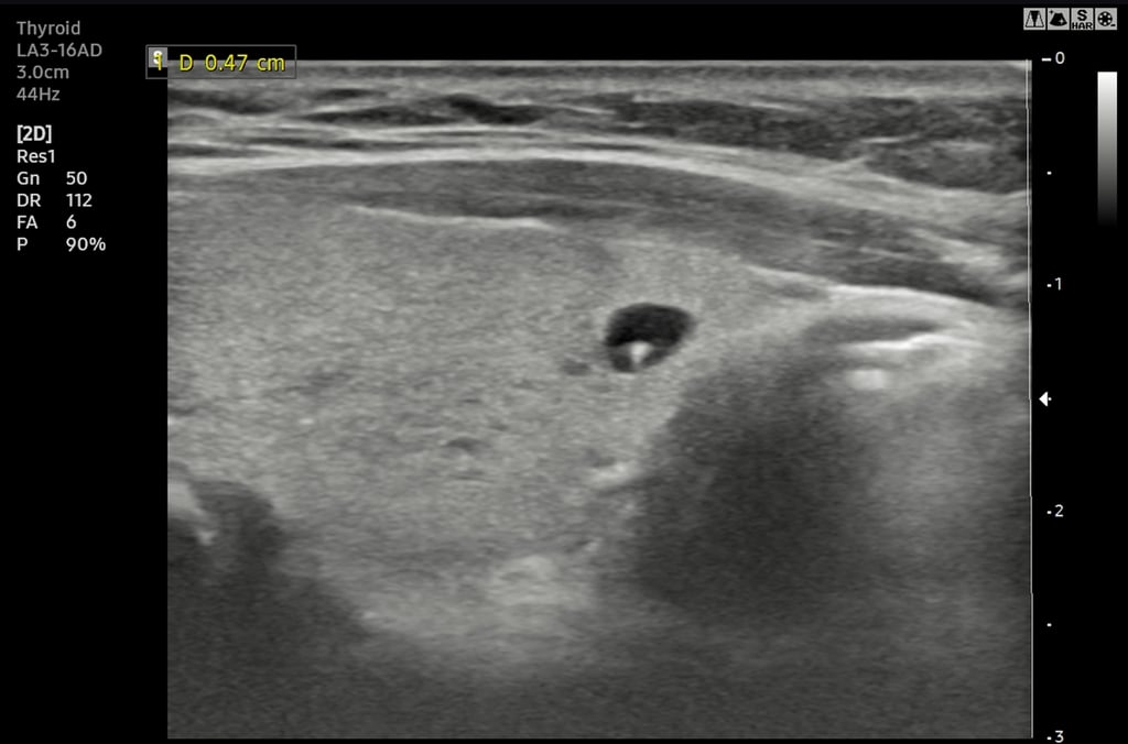

Picture 2:
Well defined thin wall small ~4mm cystic nodule with comet tail artifact, normal surrounding thyroid tissue TR1.
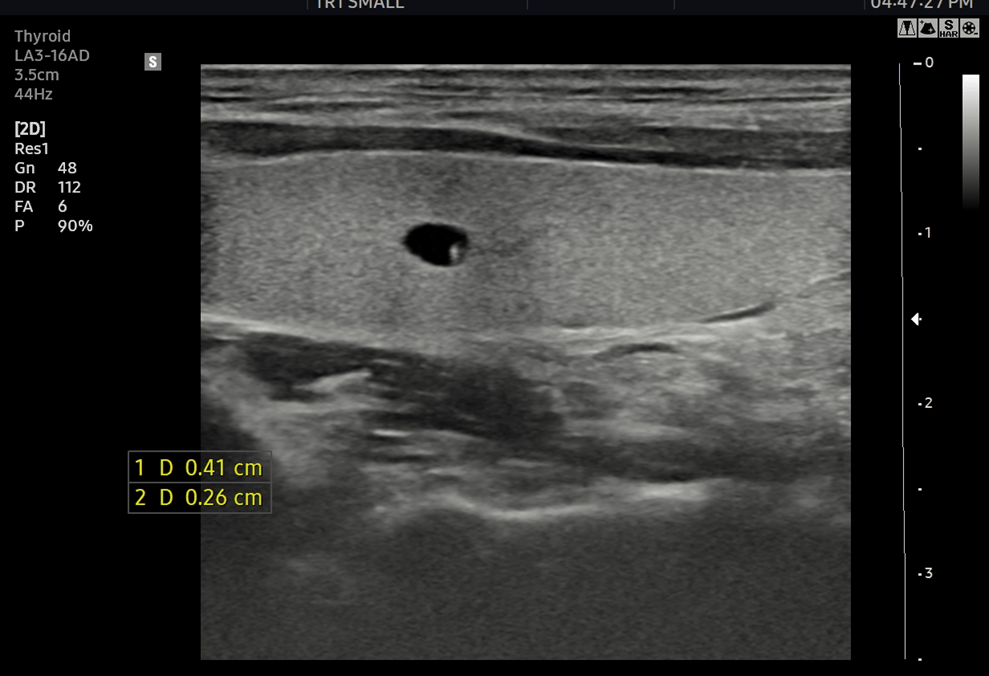

Picture 3:
Well defined thin wall small ~4mm cystic nodule with comet tail artifact, normal surrounding thyroid tissue TR1.
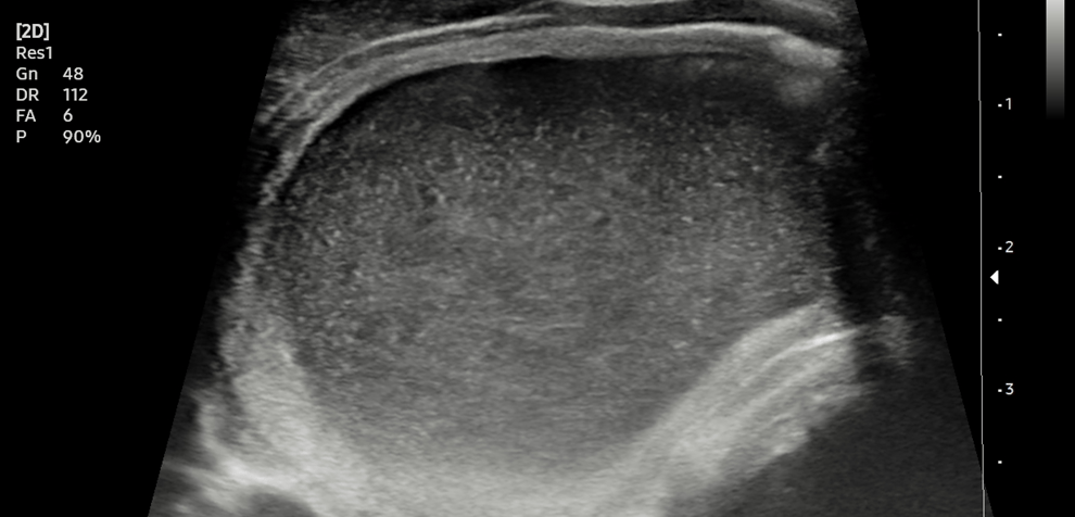

Picture 4:
Well defined thin wall cystic nodule with internal echo and fluid debris level TR1.
Picture 1:
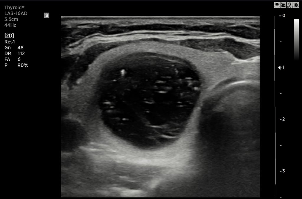

Well defined thin wall cystic nodule with internal debris and comet tail artifact.
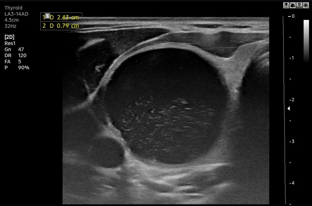

Picture 6:
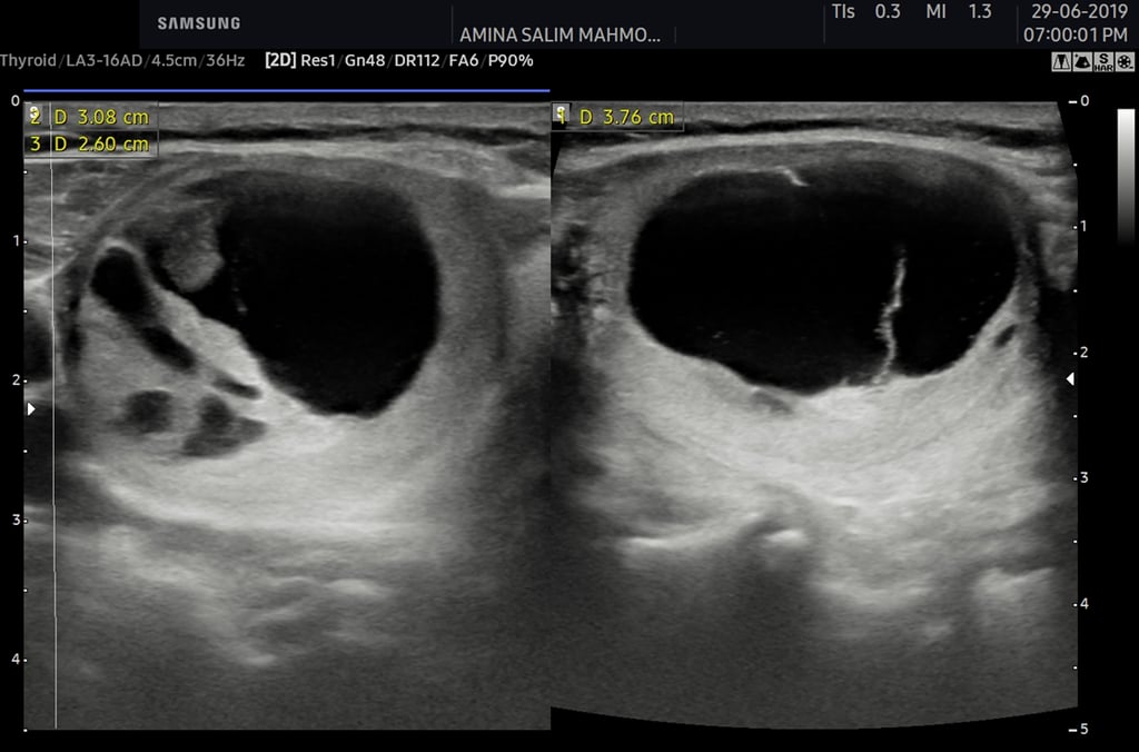

Picture 7:
Well defined complex mainly cystic nodule in Rt lobe TR2 ( solid part isoechoic without micro or macrocalcification).

Picture 8:
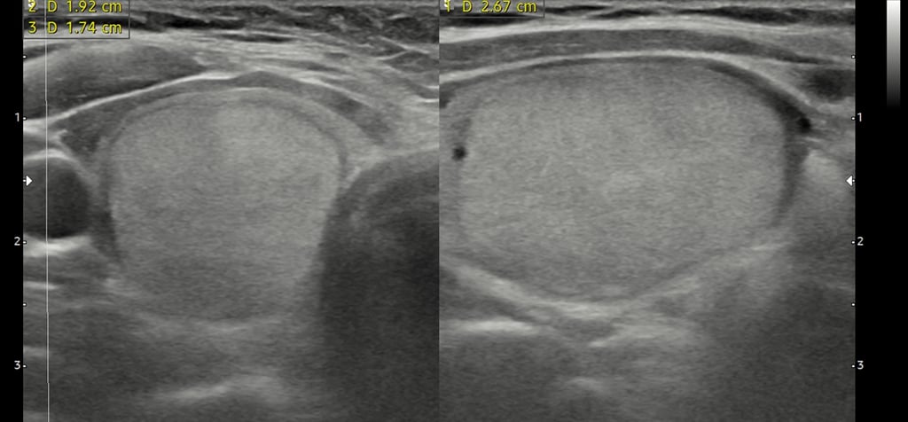
Well define smooth regular surface solid isoechoic to thyroid tissue nodule in Rt lobe low third without micro or macrocalcification TR3.
Picture 5:
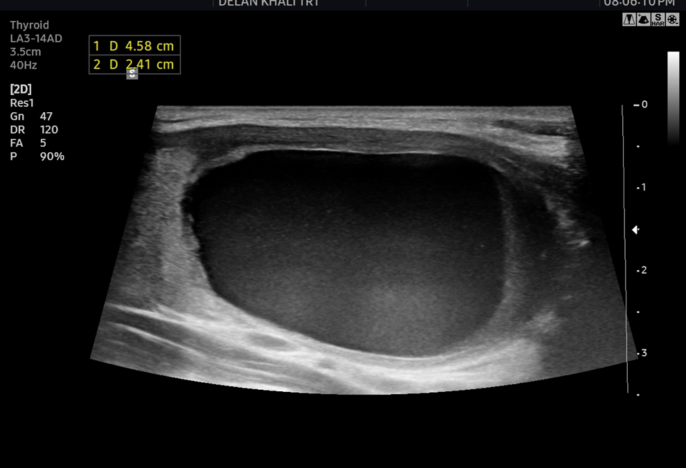

Well defined thin wall 45*24 22mm cystic nodule TR1.
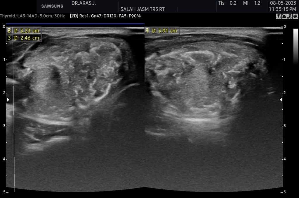

Picture 10:
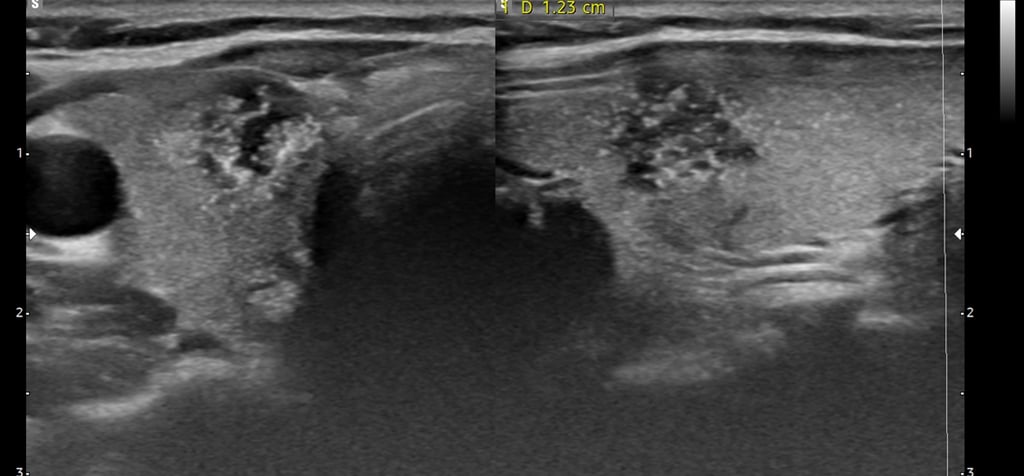
Solid hypoechoic nodule with lobulated surface , micro and macrocalcification TR5.
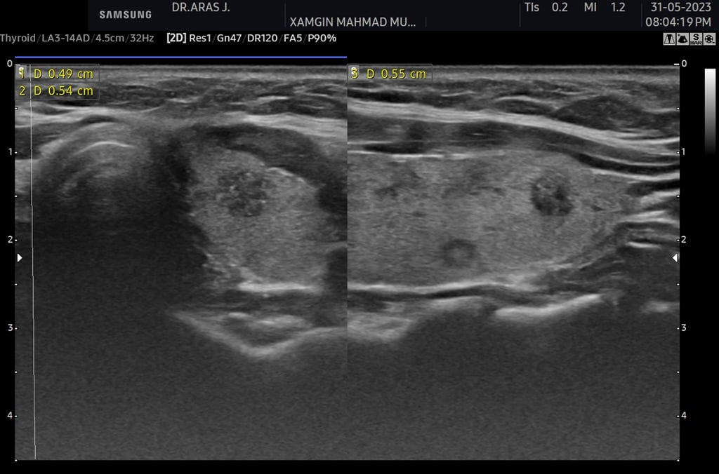

Picture 11:
Small solid lobulated surface hypoechoic nodule with microcalcification TR5.

Picture 12:
Picture 9:
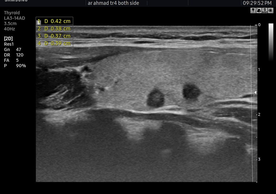

Well defined solid hypoechoic TR4 nodule.
