Tuberculous Granulomatous Inflammation of Parathyroid Adenoma Manifested as Primary Hyperparathyroidism
A 58-year-old female with a history of recurrent renal calculi presented to the Head and Neck clinic at Smart Health Tower (Sulaimani, Iraq) with complaints of generalized body aches and fatigue for approximately one year. The patient had no notable surgical history or prior infection with tuberculosis (TB).
HISTOPATHOLOGY
8/31/20231 min read


Patient Information:
A 58-year-old female with a history of recurrent renal calculi presented to the Head and Neck clinic at Smart Health Tower (Sulaimani, Iraq) with complaints of generalized body aches and fatigue for approximately one year. The patient had no notable surgical history or prior infection with tuberculosis (TB).
Clinical Findings:
Physical examination revealed no significant findings, and there was no associated palpable cervical lymphadenopathy.
Diagnostic Assessment:
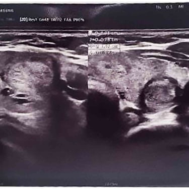
Blood analyses revealed elevated parathyroid hormone (PTH) levels (154.7 pg/ml) and serum calcium levels (11.26 mg/dl). A neck ultrasound revealed a multinodular goiter with mildly suspicious (TR3) bilateral homogeneous echo texture nodules measuring 4 mm in the right thyroid gland. The left thyroid gland had a non-suspicious (TR2) nodule measuring 13x9x8 mm and a mildly suspicious (TR3) nodule measuring 10x9x7 mm. Additionally, a solid hypoechoic hypovascular nodule of 20x7 mm below the left lower pole of the thyroid was identified, which suggested a parathyroid adenoma. No notable cervical lymphadenopathy was observed.
Therapeutic Intervention:
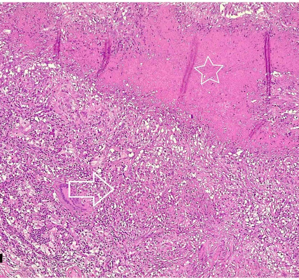
The patient underwent left thyroid lobectomy and excision of the left parathyroid gland. Histopathological examination revealed a parathyroid adenoma with caseating granulomatous inflammation, suggestive of TB. The left thyroid gland exhibited a nodular goiter with focal lymphocytic thyroiditis.
The histopathological analysis was performed using 4-µm-thick paraffin-embedded sections fixed with 10% neutral buffered formalin for 24 hours, followed by staining with hematoxylin and eosin and examination under a light microscope.
Follow-Up:
The postoperative period was uneventful, and the patient's calcium level decreased to 10 mg/dl. A negative acid-fast bacillus (AFB) test of sputum, along with negative chest X-ray findings, were achieved postoperatively. The patient was subsequently treated for TB with Rifampin (600 mg, twice daily for 6 months). The patient's symptoms, including generalized body aches, resolved following treatment.
Image Gallery


