Vascular Malformation Mimicking Parathyroid Adenoma in a 16-Year-Old Female with Elevated PTH
A 16-year-old female presented with a one-year history of anterior neck swelling associated with intermittent pain. She denied compressive symptoms such as dysphagia, dyspnea, or voice changes. Her past medical and surgical history was unremarkable, and she was not taking any chronic medications. The absence of systemic symptoms or endocrine complaints made the clinical picture initially nonspecific, though the chronicity suggested a slow-growing structural lesion.
SURGERYHEAD AND NECKVIDEO
12/2/20252 min read

Patient Presentation:
A 16-year-old female presented with a one-year history of anterior neck swelling associated with intermittent pain. She denied compressive symptoms such as dysphagia, dyspnea, or voice changes. Her past medical and surgical history was unremarkable, and she was not taking any chronic medications. The absence of systemic symptoms or endocrine complaints made the clinical picture initially nonspecific, though the chronicity suggested a slow-growing structural lesion.
Laboratory Investigations:
Laboratory evaluation revealed normal thyroid function tests, indicating a euthyroid state. However, the patient had a markedly elevated parathyroid hormone (PTH) level at 183 pg/mL (normal 15–65 pg/mL), raising suspicion for primary hyperparathyroidism. Notably, her serum calcium was normal at 9.17 mg/dL, creating a discordant biochemical profile. This pattern—high PTH with normocalcemia—can represent early primary hyperparathyroidism, secondary hyperparathyroidism, laboratory variability, or a non-parathyroid PTH-secreting lesion. This discrepancy should have triggered critical reevaluation, particularly since the imaging findings later deviated from those expected for a classic parathyroid adenoma.
Ultrasound Findings:
Thyroid ultrasonography showed right and left lobes measuring 48 × 17 × 15 mm and 46 × 15 × 14 mm respectively, with a 3 mm isthmus and overall homogeneous echotexture. The left lobe contained a TR3 nodule measuring 12 × 10 × 9 mm in the upper third and two micronodules in the mid-third. More importantly, a well-defined, lobulated, solid, heterogeneously hypoechoic mass measuring 49 × 16 × 12 mm was identified posterior to the mid-upper right thyroid lobe, exhibiting mild internal vascularity with a low-flow pattern. Sonographic interpretation included parathyroid lesion versus vascular malformation/hemangioma, with an indistinct upper pole relationship to the larynx. The size, lobulated contour, and low-flow vascular pattern raised early suspicion for a vascular lesion rather than a parathyroid adenoma.
CT Neck and Chest Imaging:
Contrast-enhanced CT demonstrated a well-defined cystic lesion measuring 19 × 14 × 13 mm supero-posterior to the right thyroid gland, containing a single internal calcific focus and limited nodular enhancement medially. Additional dense calcifications were noted along the cervical esophageal wall. Two elongated, centrally enhancing soft-tissue lesions were also observed in the right posterior cervical triangle, the largest measuring 5 × 2.5 cm. Collectively, these findings were most consistent with a posterior cervical vascular malformation with phleboliths, potentially venous or venolymphatic in nature, challenging the earlier assumption of a parathyroid adenoma based solely on elevated PTH levels.
Preoperative Assessment and Surgical Plan:
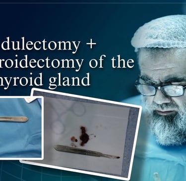
Despite the imaging favoring a vascular malformation, the elevated PTH level maintained clinical concern for parathyroid disease. Thus, the patient was scheduled for right-sided parathyroidectomy targeting both upper and lower glands, with recognition that the upper lesion could represent a hemangioma rather than parathyroid tissue. A left thyroid nodulectomy was also planned for the TR3 nodule. Preoperative assessments were within normal limits. While the surgical decision was guided by biochemical abnormality, the mismatch between imaging and expected parathyroid pathology suggested the need for additional preoperative functional imaging such as sestamibi scanning or Doppler/angiographic evaluation.
Histopathological Examination:
Histopathology of the left thyroid nodulectomy revealed a hyperplastic follicular nodule with oncocytic (Hürthle cell) changes and no malignancy. The right-sided specimen demonstrated a vascular malformation containing a single small, normal parathyroid gland. No malignancy was identified in any specimen. These findings confirmed that the mass initially interpreted as possible parathyroid pathology was instead a vascular lesion, fully consistent with CT features but discordant with the preoperative biochemical interpretation.
Final Diagnosis:
The final pathological diagnosis included:
Left thyroid hyperplastic follicular nodule with oncocytic changes — benign.
Right-sided cervical vascular malformation with incidentally included normal parathyroid gland — benign.
This case underscores the importance of correlating laboratory abnormalities with imaging patterns and recognizing that elevated PTH alone should not override contradictory anatomic evidence, particularly when imaging strongly suggests vascular pathology.

Address
Majid Bag Main Street, Beside University of Sulaymaniyah Old Campus, Madam Mitterrand, Sulaymaniyah, Iraq
Contacts
info@ssthyroid.com