Vascular Malformation of the Right Nasal Wall and Cheek
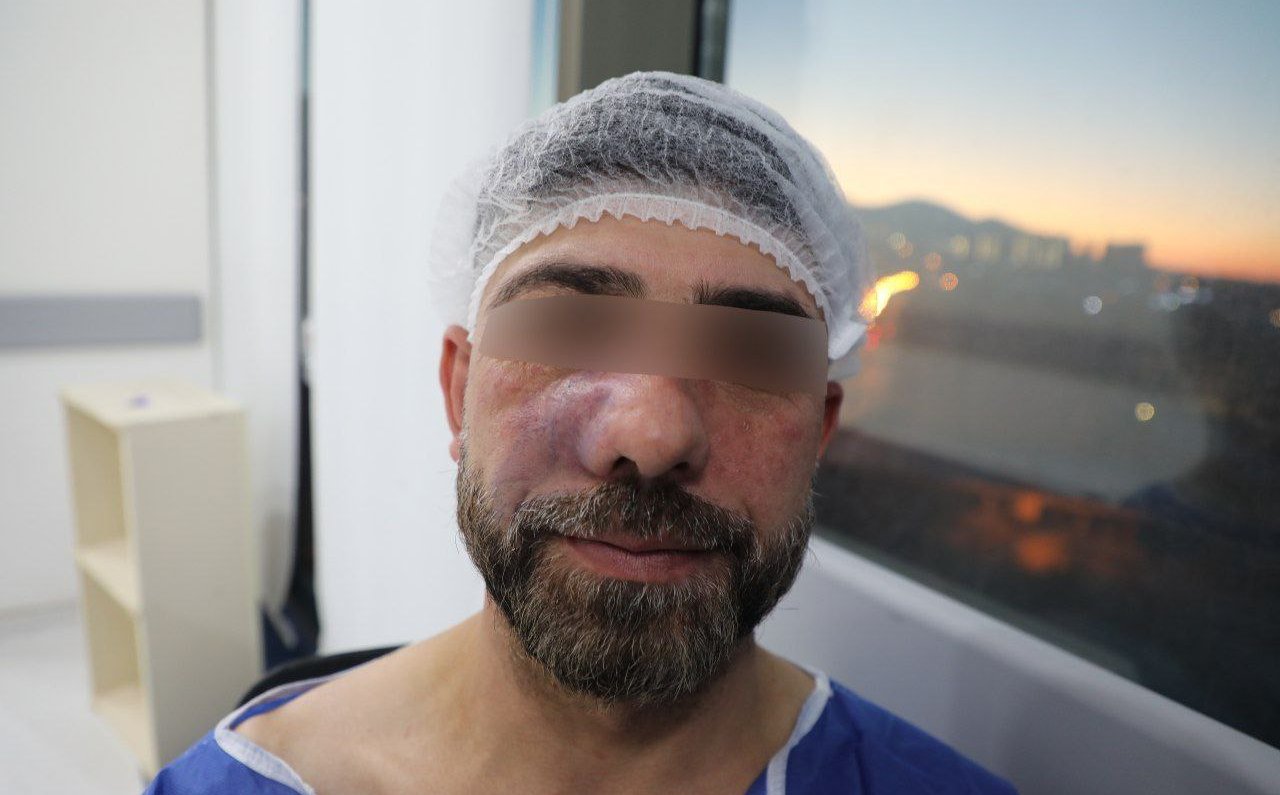
A 41-year-old male presented with a long-standing lesion on the right side of his nose. The patient has a history of smoking, but no notable past medical or surgical history. He has previously undergone sclerotherapy two years ago, which failed to resolve the issue.
SURGERYHEAD AND NECKVIDEO
2/20/20251 min read


Patient Information:
A 41-year-old male presented with a long-standing lesion on the right side of his nose. The patient has a history of smoking, but no notable past medical or surgical history. He has previously undergone sclerotherapy two years ago, which failed to resolve the issue.
Clinical Findings:
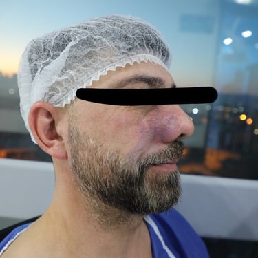
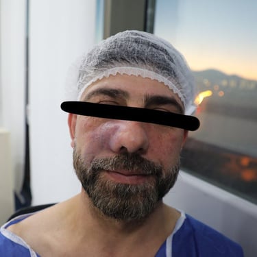
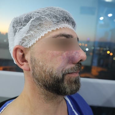
The patient has a vascular malformation on the right nasal wall and cheek. The lesion involves the skin and subcutaneous tissue, with a complex heterogeneous texture on ultrasound. The lesion shows multiple low-flow vessels (<2mm in diameter), suggestive of a hemangioma. CT scan indicated internal extension into the nose. Despite having been scheduled for sclerotherapy abroad, the consultant believes that the procedure will likely fail due to the diffuse nature and multiple feeders of the malformation.
Diagnostic Assessment:
Ultrasound of the neck and face revealed a lesion 50x44x18mm in size, involving both the cheek and nose.
No pathological lymphadenopathy or issues with the thyroid were observed.
Blood tests and ECG were normal, and viral screening was negative.
Therapeutic Intervention:
Due to the lesion's diffuse nature and involvement with the skin, surgical resection was planned. A biopsy and mesh fixation were performed, confirming the diagnosis of a hemangioma vascular malformation through histopathological examination (HPE).
Follow-up:
Post-operative care included monitoring for complications, and further treatment was planned based on the outcome of the resection. A plastic surgeon may be consulted for optimal aesthetic outcomes given the involvement of the skin.
Gallery


