Vascular Malformation with Discordant Ultrasound Features in a 12-Year-Old Boy
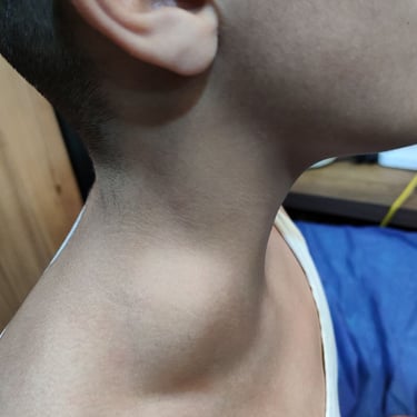
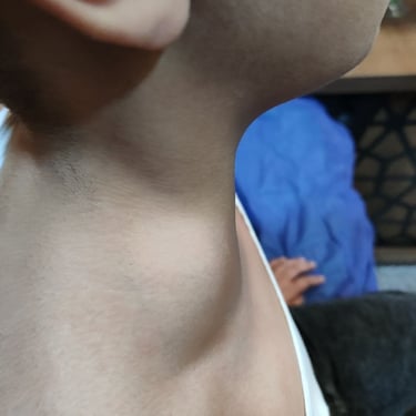
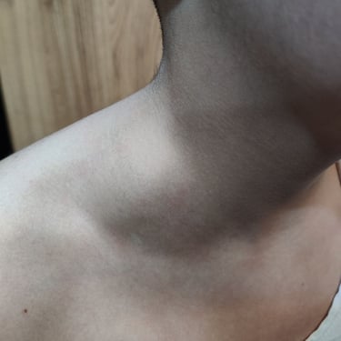
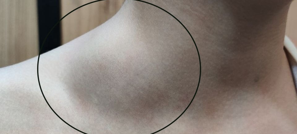
A 12-year-old boy presented with a right-sided neck swelling that had developed over the past 10 days. He had no significant past medical, surgical, or drug history. The swelling was non-tender and had no associated systemic symptoms such as fever, pain, or respiratory distress.
HEAD AND NECK
1/11/20241 min read

Case Presentation:
A 12-year-old boy presented with a right-sided neck swelling that had developed over the past 10 days. He had no significant past medical, surgical, or drug history. The swelling was non-tender and had no associated systemic symptoms such as fever, pain, or respiratory distress.
Laboratory Findings:
All laboratory parameters were within normal ranges, including a complete blood count (CBC), erythrocyte sedimentation rate (ESR: 6 mm/hr), thyroid-stimulating hormone (TSH: 2.15 uIU/ml), and free thyroxine (FT4: 16.36 pmol/L).
Imaging Findings:
Neck ultrasound revealed multiple bilateral cervical lymph nodes, predominantly on the right side, with the largest lymph node (28×8 mm) in the right submandibular region, suggesting an inflammatory response. Additionally, a well-defined, thin-walled (~1mm) cystic lesion measuring 62×44×19 mm was identified in the right supraclavicular region. The lesion was hypovascular, had no solid component or internal septations, and was suggestive of a cystic hygroma or, less likely, a lymphangioma. The thyroid, submandibular, and parotid glands were normal, with no focal lesions.
Management Plan:
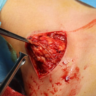
Following a multidisciplinary team (MDT) discussion, surgical excision of the cystic mass was recommended to obtain a definitive diagnosis and guide further treatment if needed. The excised specimen was sent for histopathological examination (HPE).
Histopathology Findings:
Histological analysis revealed fibroadipose tissue containing thin- and thick-walled anastomosing venous, arterial, and lymphatic vascular spaces lined by benign endothelial cells. The presence of congestion, mild inflammation, and multifocal brown adipocytes confirmed the diagnosis of a macrocystic vascular malformation.
Conclusion:
The case highlights a vascular malformation with atypical ultrasound findings. The final diagnosis was established through histopathological evaluation, which distinguished the lesion from other possible differentials such as lymphangioma. Given the benign nature of the lesion, further management will depend on clinical follow-up and symptom progression.
Image Gallery
