تخطي إلى المحتوى
Smart Surgical Team
المنشورات
المحتوىالبحوثالملصقات
الأخبارالفعاليات
حول
من نحناتصل بنا
Englishالعربية
تسجيل الدخولإنشاء حساب
المنشورات
المحتوىالبحوثالملصقات
الأخبارالفعاليات
حول
من نحناتصل بنا
اللغة
Englishالعربية
تسجيل الدخولإنشاء حساب
المحتوى//Tender Left Thyroid Swelling Due to Hemorrhagic Cyst in a Hyperplastic Nodule: A Benign Mimic of Thyroid Malignancy
مقالة حالة سريرية · Clinical education

Tender Left Thyroid Swelling Due to Hemorrhagic Cyst in a Hyperplastic Nodule: A Benign Mimic of Thyroid Malignancy

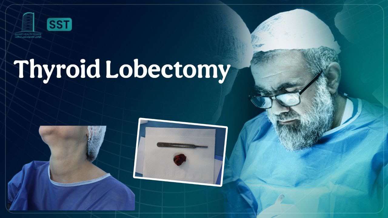
A 45-year-old female presented with a complaint of neck swelling and tenderness, predominantly on the left side. The onset was recent and acute in character. She denied dysphagia, voice changes, systemic symptoms, or history of trauma. There was no significant past medical or surgical history. Clinical examination revealed an asymmetric enlargement of the left thyroid lobe with localized tenderness, without palpable lymphadenopathy.
المشاهدة على YouTubeيفتح في YouTube للمحتوى المقيّد بحسب العمر
Smart Surgical Team
LinkedIn

روابط سريعة

المحتوىالبحوثالملصقاتالفعالياتمن نحناتصل بنا

تصفّح حسب الموضوع

الغدة الدرقية وجارات الدرقيةالغدد اللعابيةجراحة العنق والجهاز اللمفاويالجلد والأنسجة الرخوة

اتصل بنا

info@smartsurgicalteam.com

شارع ماجد باغ الرئيسي، بجوار الحرم القديم لجامعة السليمانية، مدام ميتران، السليمانية، العراق

© 2026 Smart Surgical Team. جميع الحقوق محفوظة.سياسة الخصوصيةشروط الاستخدام

تفاصيل الحالة

مقالة حالة سريرية
العرض السريري
A 45-year-old female presented with a complaint of neck swelling and tenderness, predominantly on the left side. The onset was recent and acute in character. She denied dysphagia, voice changes, systemic symptoms, or history of trauma. There was no significant past medical or surgical history. Clinical examination revealed an asymmetric enlargement of the left thyroid lobe with localized tenderness, without palpable lymphadenopathy.
التصوير والاستقصاءات

Thyroid function tests were within normal limits:

  • TSH: 1.41 uIU/mL

  • Free T4: 14.9 pmol/L

Inflammatory markers were significantly elevated:

  • Erythrocyte Sedimentation Rate (ESR): 40 mm/hr

  • C-reactive Protein (CRP): 55.9 mg/L

Other relevant labs:

  • Parathyroid Hormone (PTH): 69.4 pg/mL (normal to mildly elevated range)

These findings raised concern for an inflammatory or hemorrhagic process involving the thyroid.

Ultrasound of the Thyroid:

  • Right lobe: 47 × 18 × 16 mm, normal in size, with a few small TR2 nodules (largest 7 × 6 × 4 mm).

  • Isthmus: ~3 mm, no focal lesions.

  • Left lobe: 45 × 37 × 32 mm, significantly enlarged with inhomogeneous echotexture.

Notable findings:

  • A well-defined, regular-surfaced, mixed (predominantly cystic) nodule measuring 37 × 30 × 27 mm occupying most of the left lobe.

  • TR2 classification with no microcalcifications, no macrocalcifications, and increased perinodular vascularity.

  • A small (approx. 4 mm thick), ill-defined hypoechoic lesion noted in the lower third of the left lobe, suspicious for surrounding hematoma.

Ultrasound impression: Large hemorrhagic cyst in a benign-appearing nodule; low suspicion for malignancy but correlation with clinical course and histology recommended.

الإجراء الجراحي
Due to the size of the nodule, associated tenderness, inflammatory markers, and the suspicion of intralesional hemorrhage, tissue diagnosis was pursued. The lesion was excised or biopsied (not explicitly mentioned, assumed to be FNA or surgical excision depending on clinical judgment).
علم الأمراض النسيجي

Final histological analysis revealed:

  • Hyperplastic thyroid follicular nodule with cystic degeneration.

  • No features of malignancy.

  • Hemorrhagic and degenerative changes consistent with the imaging and clinical picture.


النتيجة والمتابعة
This case illustrates a benign yet symptomatic thyroid nodule with cystic and hemorrhagic changes, mimicking features of malignancy both clinically and radiologically. The elevated inflammatory markers and rapid onset raised concern, but histological analysis confirmed a hyperplastic nodule without malignancy. This underscores the importance of tissue diagnosis in atypical or symptomatic thyroid lesions to avoid overtreatment and misdiagnosis.
المساهم
صورة Prof. Abdulwahid M. Salih

Prof. Abdulwahid M. Salih

M.D. (General Surgery)

عرض الفريق
تفاصيل المحتوى
الصيغة
مقالة حالة سريرية
الموضوع
Thyroid Nodules & Cysts
المستوى
Clinical education
صور الحالة

محتوى ذو صلة

عرض المكتبة

قبلبعد
Thyroid & Parathyroid

Targeted Parathyroidectomy for Incidental Parathyroid Adenoma in a 10-Year-Old Female

قبلبعد
Skin & soft-tissue

Submental Dermoid Cyst in an 15 year old Female

قبلبعد
Thyroid & Parathyroid

Advanced Papillary Thyroid Carcinoma with Nodal and Distant Spread