Skip to content
Smart Surgical Team
Publications
ContentResearchPosters
NewsEvents
About
About usContact
Englishالعربية
Sign inRegister
Publications
ContentResearchPosters
NewsEvents
About
About usContact
Language
Englishالعربية
Sign inRegister
Content//Tender Left Thyroid Swelling Due to Hemorrhagic Cyst in a Hyperplastic Nodule: A Benign Mimic of Thyroid Malignancy
Case article · Clinical education

Tender Left Thyroid Swelling Due to Hemorrhagic Cyst in a Hyperplastic Nodule: A Benign Mimic of Thyroid Malignancy

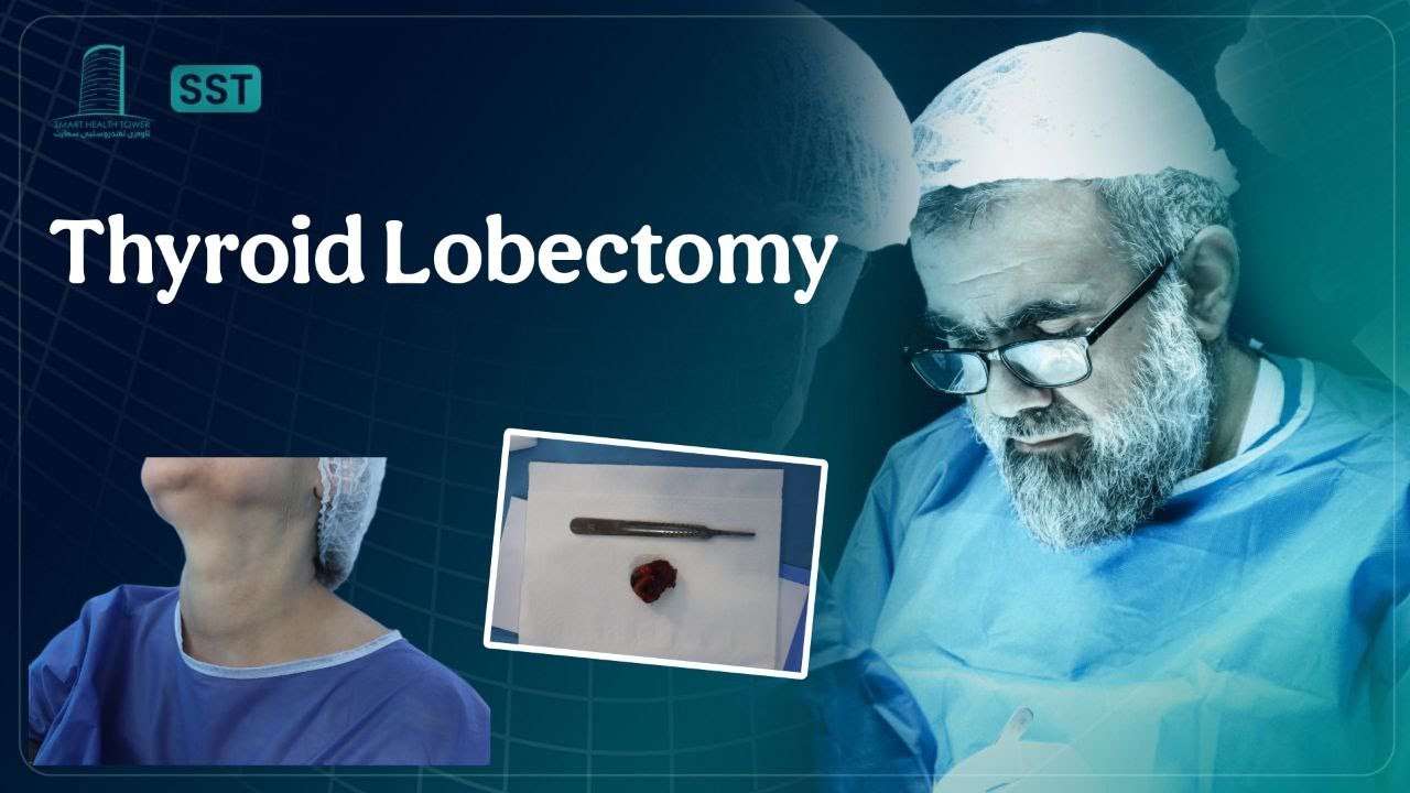
A 45-year-old female presented with a complaint of neck swelling and tenderness, predominantly on the left side. The onset was recent and acute in character. She denied dysphagia, voice changes, systemic symptoms, or history of trauma. There was no significant past medical or surgical history. Clinical examination revealed an asymmetric enlargement of the left thyroid lobe with localized tenderness, without palpable lymphadenopathy.
Watch on YouTubeOpens in YouTube for age-restricted content
Smart Surgical Team
LinkedIn

Quick links

ContentResearchPostersEventsAbout usContact us

Browse by Topic

Thyroid & ParathyroidSalivary GlandsNeck & Lymphatic SurgerySkin & Soft Tissue

Contact us

info@smartsurgicalteam.com

Majid Bag Main Street, Beside University of Sulaymaniyah Old Campus, Madam Mitterrand, Sulaymaniyah, Iraq

© 2026 Smart Surgical Team. All rights reserved.Privacy PolicyTerms of Use

Case details

Case article
Presentation
A 45-year-old female presented with a complaint of neck swelling and tenderness, predominantly on the left side. The onset was recent and acute in character. She denied dysphagia, voice changes, systemic symptoms, or history of trauma. There was no significant past medical or surgical history. Clinical examination revealed an asymmetric enlargement of the left thyroid lobe with localized tenderness, without palpable lymphadenopathy.
Imaging & workup

Thyroid function tests were within normal limits:

  • TSH: 1.41 uIU/mL

  • Free T4: 14.9 pmol/L

Inflammatory markers were significantly elevated:

  • Erythrocyte Sedimentation Rate (ESR): 40 mm/hr

  • C-reactive Protein (CRP): 55.9 mg/L

Other relevant labs:

  • Parathyroid Hormone (PTH): 69.4 pg/mL (normal to mildly elevated range)

These findings raised concern for an inflammatory or hemorrhagic process involving the thyroid.

Ultrasound of the Thyroid:

  • Right lobe: 47 × 18 × 16 mm, normal in size, with a few small TR2 nodules (largest 7 × 6 × 4 mm).

  • Isthmus: ~3 mm, no focal lesions.

  • Left lobe: 45 × 37 × 32 mm, significantly enlarged with inhomogeneous echotexture.

Notable findings:

  • A well-defined, regular-surfaced, mixed (predominantly cystic) nodule measuring 37 × 30 × 27 mm occupying most of the left lobe.

  • TR2 classification with no microcalcifications, no macrocalcifications, and increased perinodular vascularity.

  • A small (approx. 4 mm thick), ill-defined hypoechoic lesion noted in the lower third of the left lobe, suspicious for surrounding hematoma.

Ultrasound impression: Large hemorrhagic cyst in a benign-appearing nodule; low suspicion for malignancy but correlation with clinical course and histology recommended.

Procedure
Due to the size of the nodule, associated tenderness, inflammatory markers, and the suspicion of intralesional hemorrhage, tissue diagnosis was pursued. The lesion was excised or biopsied (not explicitly mentioned, assumed to be FNA or surgical excision depending on clinical judgment).
Histopathology

Final histological analysis revealed:

  • Hyperplastic thyroid follicular nodule with cystic degeneration.

  • No features of malignancy.

  • Hemorrhagic and degenerative changes consistent with the imaging and clinical picture.


Outcome & follow-up
This case illustrates a benign yet symptomatic thyroid nodule with cystic and hemorrhagic changes, mimicking features of malignancy both clinically and radiologically. The elevated inflammatory markers and rapid onset raised concern, but histological analysis confirmed a hyperplastic nodule without malignancy. This underscores the importance of tissue diagnosis in atypical or symptomatic thyroid lesions to avoid overtreatment and misdiagnosis.
Contributor
Portrait of Prof. Abdulwahid M. Salih

Prof. Abdulwahid M. Salih

M.D. (General Surgery)

View team
Content details
Format
Case article
Topic
Thyroid Nodules & Cysts
Level
Clinical education
Case images

Related content

View library

BeforeAfter
Thyroid & Parathyroid

Targeted Parathyroidectomy for Incidental Parathyroid Adenoma in a 10-Year-Old Female

BeforeAfter
Skin & soft-tissue

Submental Dermoid Cyst in an 15 year old Female

BeforeAfter
Thyroid & Parathyroid

Advanced Papillary Thyroid Carcinoma with Nodal and Distant Spread