Skip to content
Smart Surgical Team
Publications
ContentResearchPosters
NewsEvents
About
About usContact
Englishالعربية
Sign inRegister
Publications
ContentResearchPosters
NewsEvents
About
About usContact
Language
Englishالعربية
Sign inRegister
Content//Recurrent Papillary Thyroid Carcinoma with Bilateral Lateral Cervical Lymph Node Metastasis
Operative video · Clinical education

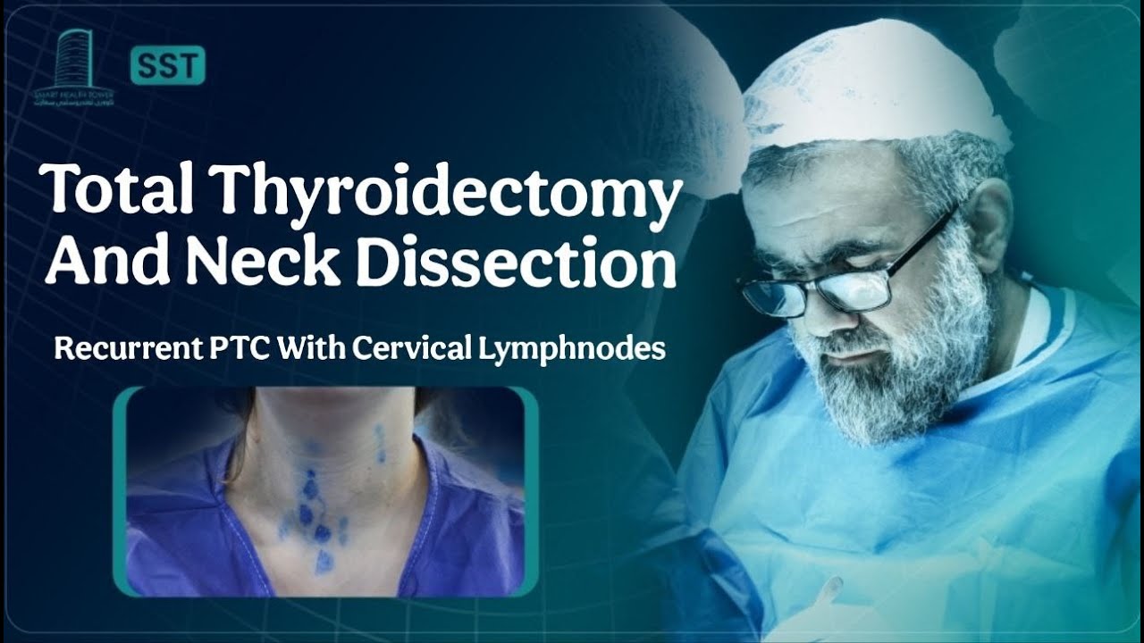
Recurrent Papillary Thyroid Carcinoma with Bilateral Lateral Cervical Lymph Node Metastasis

A 35-year-old female presented with a history of thyroid disease of 3 years duration. She had undergone a thyroid operation three years prior for papillary thyroid carcinoma and had received radioactive iodine (RAI) therapy postoperatively. She reported no significant new symptoms at the time of presentation.
Watch on YouTubeOpens in YouTube for age-restricted content
Smart Surgical Team
LinkedIn

Quick links

ContentResearchPostersEventsAbout usContact us

Browse by Topic

Thyroid & ParathyroidSalivary GlandsNeck & Lymphatic SurgerySkin & Soft Tissue

Contact us

info@smartsurgicalteam.com

Majid Bag Main Street, Beside University of Sulaymaniyah Old Campus, Madam Mitterrand, Sulaymaniyah, Iraq

© 2026 Smart Surgical Team. All rights reserved.Privacy PolicyTerms of Use

Case details

Operative video
Presentation
A 35-year-old female presented with a history of thyroid disease of 3 years duration. She had undergone a thyroid operation three years prior for papillary thyroid carcinoma and had received radioactive iodine (RAI) therapy postoperatively. She reported no significant new symptoms at the time of presentation.
Imaging & workup

Investigations:
Laboratory tests revealed elevated Thyroid-Stimulating Hormone (TSH) at 49.8 uIU/ml and low-normal Free T4 at 10.3 pmol/L, indicating hypothyroidism. Serum calcium was 9.18 mg/dl and parathyroid hormone (PTH) level was 35.5 pg/ml, within normal limits. Calcitonin was <0.500 pg/ml. Thyroglobulin was elevated at 62.6 ng/ml. Renal function tests and complete blood count were within normal limits, and viral screening was negative. Vocal cord examination showed normal mobility, shape, and color of both vocal cords.

Neck Ultrasound Findings:
Ultrasound of the neck showed remnant thyroid tissue in the right lobe operation bed measuring approximately 39 × 18 × 17 mm with inhomogeneous echotexture and multiple solid, hypoechoic, vascular nodules with microcalcifications, consistent with highly suspicious TR5 nodules or metastatic lymph nodes. Similar nodules were noted in the left thyroid bed and in the midline over the trachea extending retrosternally. Additionally, multiple suspicious cervical lymph nodes were identified bilaterally, with microcalcifications and loss of normal hilar architecture. The largest right-sided LN was 16 × 5 mm (group III), and the largest left-sided LN was 12 × 3 mm (group II). Submandibular and parotid glands appeared normal.

Fine Needle Aspiration Results:
FNA of the right TR5 nodule and bilateral cervical lymph nodes confirmed recurrence of papillary thyroid carcinoma. The right thyroid remnant was Bethesda category VI, and both right and left cervical lymph nodes tested positive for metastatic papillary thyroid carcinoma.

Procedure
Given the findings, the patient was scheduled for revision thyroid surgery. She underwent recurrent thyroidectomy with bilateral central lymph node dissection (CLND) and bilateral lateral lymph node dissection (LLND). Under general anesthesia, a collar incision was made in the supine, neck-extended position. Total thyroidectomy was performed, preserving both recurrent laryngeal nerves and parathyroid glands. Redivac drains were placed, and the wound was closed in layers. Dissection included bilateral lateral neck groups (levels II–V) and bilateral central compartments. All major vessels and nerves, including the vagus and phrenic nerves, were preserved. The procedure duration was 4 hours and 30 minutes.
Histopathology

Histological examination confirmed metastatic papillary thyroid carcinoma involving 18 of 65 lymph nodes. Specifically:

  • Right central group: 11 of 11 nodes involved.

  • Left central group: 4 of 5 nodes involved.

  • Right lateral group: 0 of 20 nodes involved.

  • Left lateral group: 3 of 29 nodes involved.
    No residual thyroid tissue was seen; only skeletal muscle and fibrous tissue were identified at the thyroid bed.

Outcome & follow-up
Postoperative Course:
The patient tolerated the procedure well with no immediate complications. She was stable postoperatively and scheduled for further oncological evaluation and follow-up.
Contributor
Portrait of Prof. Abdulwahid M. Salih

Prof. Abdulwahid M. Salih

M.D. (General Surgery)

View team
Content details
Format
Operative video
Topic
Papillary Thyroid Carcinoma
Level
Clinical education
Case images

Related content

View library

Sialolithiasis & Sialadenitis

Right Submandibular Sialolithiasis with Non-Specific Sialadenitis

Salivary Glands

Recurrent Multifocal Pleomorphic Adenoma of the Right Parotid Gland in a 37-Year-Old Male

Thyroid & Parathyroid

Massive Multinodular Goiter With Retrosternal Extension in a Patient With Long-Standing Thyroid Disease