Operative video · Clinical case
Vascular Malformation Mimicking Parathyroid Adenoma in a 16-Year-Old Female with Elevated PTH
A cervical vascular malformation mimicked parathyroid adenoma in a 16-year-old with elevated PTH but normal calcium.
A cervical vascular malformation mimicked parathyroid adenoma in a 16-year-old with elevated PTH but normal calcium.
A 16-year-old female presented with a one-year history of anterior neck swelling and intermittent pain, without dysphagia, dyspnoea or voice change. Her medical and surgical history was unremarkable.
Thyroid function was normal, but PTH was 183 pg/mL with normal calcium (9.17 mg/dL). Ultrasound showed a 49 × 16 × 12 mm lobulated posterior right-thyroid mass with low-flow vascularity, considered parathyroid lesion versus vascular malformation. CT demonstrated a cystic superoposterior right-thyroid lesion with calcification plus enhancing posterior-triangle lesions, features most consistent with a posterior cervical vascular malformation with phleboliths.
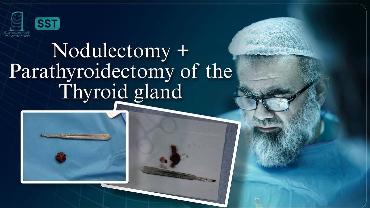
A right-sided parathyroid exploration targeting upper and lower glands was planned because of the biochemical concern, with concurrent left thyroid nodulectomy for a TR3 nodule.
The left nodule was a benign hyperplastic follicular nodule with oncocytic change. The right-sided specimen was a vascular malformation containing a small normal parathyroid gland; no malignancy was found.
The final diagnosis was benign cervical vascular malformation rather than parathyroid adenoma, emphasising the need to reconcile biochemical results with contradictory imaging.